Kentaro Wakamatsu, MD, PhD1, Zenzo Nagasawa, MT, PhD2, Kouta Katsuki, MT 3, Hiroyuki Kumazoe, MD1, Masayo Yasuda, MT 3, Sae Kawamoto, MT 3, Ayano Kawamura, MT 3, Junji Otsuka, M.D, PhD1, Ruriko Kiyotani1, Izumi Fukui1, Sanae Maki, MD, PhD1, Nobuhiko Nagata, MD, PhD4, Masayuki Kawasaki, MD, PhD1, Satomi Asai, MD, PhD5, Hozumi Yamada, MD, PhD6
Wakamatsu K, Nagasawa Z, Katsuki K, Kumazoe H, Yasuda M, Kawamoto S, Kawamura A, Otsuka J, Kiyotani R, i Fukui I, Maki S, Nagata N, Kawasaki M, Asai S, Yamada H. Retrospective study of the prognosis, viral load estimated by RT-PCR cycle threshold values, and monocyte distribution width (MDW) in COVID-19. Lab Med Int 2024; 3(3): 84-94. doi: 10.51041/lmi.3.3_84
Original
Lab Med Int 2024; 3(3): 84-94
†Correspondence: Department of Respiratory Medicine, National Hospital Organization Omuta National Hospital
1044-1 Oaza, Tachibana, Omuta City, Fukuoka 837-0911, Japan
E-mail: wakamatsu.kentaro.fe”@”mail.hosp.go.jp
Received October 3, 2023; accepted May 14, 2024
*1 Department of Respiratory Medicine, National Hospital Organization Omuta National Hospital
*2 Department of Medical Technology and Science, Faculty of Fukuoka Health Care, International University of Health and Welfare
*3 Department of Clinical Laboratory, National Hospital Organization Omuta National Hospital
*4 Department of Respiratory Medicine, Fukuoka Sanno Hospital
*5 Department of Laboratory Medicine, Tokai University School of Medicine
*6 Department of Respiratory Medicine, Keitendo Koga Hospital
ABSTRACT
Objectives: There are reports on prognostic factors including viral load, the presence/absence of pneumonia and MDW at the time of hospital admission, however there are several challenges.
Methods:
Cohort 1: A total of 235 patients diagnosed with COVID-19 admitted to our hospital between July 2020 and July 2021 were investigated for prognostic factors including the presence/absence of concurrent pneumonia and viral load.
Cohort 2: This investigation planned to investigate the prognostic significance of MDW included 80 patients diagnosed with COVID-19 who were admitted to our hospital between April 2021 when automatic blood cell counter measuring MDW was introduced in our hospital and September 2021.
Results:
Cohort 1: Based on the results of multivariate analysis, advanced age, high body mass index (BMI), diabetes mellitus, and concurrent pneumonia were considered to be independent poor prognostic factors.
Cohort 2: The mean age and MDW on admission were significantly higher in the poor prognosis group (n=38) than in the good prognosis group (n=42) (p<0.01), while high BMI, diabetes mellitus, and concurrent pneumonia showed no significant difference between the two groups.
Conclusions: MDW may be used as a biomarker of aggravation of COVID-19 on admission.
〔Lab Med Int 2024; 3(3): 84-94〕
Key Words
COVID-19, SARS-CoV-2, MDW, Ct, Prognosis
List of abbreviations
Alb=albumin; ALT=alanine aminotransferase; APTT=activated partial thromboplastin time; AUC=area under the curve; AST=aspartate aminotransferase; BMI=body mass index; BUN=blood urea nitrogen; CBC=complete blood count; CDC=Center for Disease Control and Prevention; CK=Creatine Kinase; COPD=chronic obstructive pulmonary disease; COVID-19 =coronavirus disease 2019; Cre=creatinine; CRP=C-reactive protein; Ct=cycle threshold; CT=Computed Tomography; EDTA=ethylenediaminetetraacetic acid; Glu=glucose; Hb=hemoglobin; HbA1c=Hemoglobin A1c; KL-6=krebs von den lungen-6; LD=lactate dehydrogenase; Ly=lymphocyte count; MDW=monocyte distribution width; Neu=Neutrophil count; NLR=neutrophil-to-lymphocyte ratio; NPV=negative predictive value; PCR=polymerase chain reaction; PCT=procalcitonin; Plt=platelet count; PPV=positive predictive value; PT=prothrombin time; RBC=red blood cell; RNA=ribo nucleic acid; ROC=receiver operating characteristic; RT-PCR=Reverse Transcription-PCR; SARS-CoV-2=severe acute respiratory syndrome coronavirus 2; SIRS=systemic inflammatory response syndrome; SP-A=surfactant protein A; SP-D=surfactant protein D; SpO2=oxygen saturation of peripheral artery; T-bil=total bilirubin; US=United States; WBC=white blood cell; WHO=World Health Organization
I.Introduction
Coronavirus disease 2019 (COVID-19) is an acute respiratory infection caused by severe acute respiratory syndrome coronavirus 2 (SARS-CoV-2) which was originally reported in December 2019 as “pneumonia of unknown cause” in Wuhan, Hubei, China. It was officially pronounced a new type of coronavirus by the Chinese Center for Disease Control and Prevention (CDC) on January 7, 2020. 1)
We often experience patients with COVID-19 not having any serious respiratory symptoms on admission, however rapid aggravation of respiratory status during follow-up. Therefore, for the treatment of COVID-19, it is important to diagnose the disease early, predict its aggravation, and provide therapeutic intervention early. The 6.2th edition of the Clinical Practice Guidelines for Novel Coronavirus Infections (COVID-19) lists the following risk factors for aggravation: (1) elderly persons aged 65 years or older, (2) malignant tumor, (3) chronic obstructive pulmonary disease (COPD), (4) chronic kidney disease, (5) type 2 diabetes mellitus, (6) hypertension, (7) dyslipidemia, (8) obesity (BMI of 30 kg/m² or higher), (9) smoking, (10) immunodeficiency after solid organ transplant, (11) late pregnancy. Multiple factors have also been listed as markers of aggravation, including (1) elevated D-dimer, (2) elevated CRP, (3) elevated LD, (4) elevated ferritin, (5) lymphopenia, (6) elevated creatinine, (7) elevated troponin, and (8) elevated KL-6. 2
Computed Tomography (CT) scans can be performed relatively easily, since our hospital has a dedicated CT examination device for COVID-19 patients. It is also possible to estimate the viral RNA amount using the number of positive Ct value of the Smart Gene®, a PCR reagent for SARS-CoV-2 detection (MIZUHO MEDY Co., Ltd.)3). There are reports on prognostic factors including viral load and the presence/absence of pneumonia at the time of hospital admission, however there are several challenges.
As previously reported, in recent years, devices that can analyze detailed white blood cell information with automated hematology analyzers have appeared, and it has been reported that Monocyte Distribution Width (MDW) is useful for diagnosing sepsis and assessing the severity of COVID-19 4)-23).
In this study we examined prognostic factors in COVID-19, including the presence/absence of concurrent pneumonia and viral load, and also examined MDW.
II.Methods
1. Study subjects
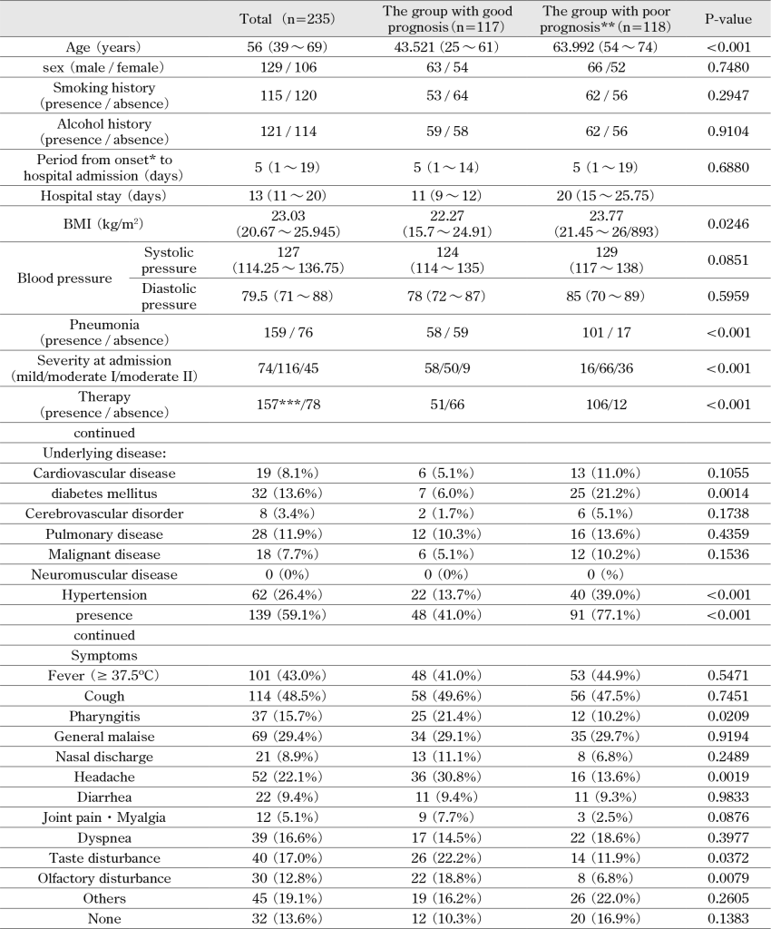
Cohort 1: The study included 235 patients (severity: mild: 74 patients, moderate I: 116 patients, and moderate II: 45 patients) admitted to the National Hospital Organization Omuta Hospital and confirmed to have COVID-19 by SARS-CoV-2 PCR test between July 2020 and July 2021. In the evaluation, a comparative evaluation was performed retrospectively using blood samples collected at the initial visit. Severity was classified according to the 6.2th edition of the Clinical Practice Guidelines2). The details of patient background are shown in Table 1.
In the diagnosis of COVID-19, nasopharyngeal swabs were collected from patients and the samples are analyzed by a fully automated genetic analyzer, Smart Gene® (Mizuho Medy, Co., Ltd.).
Pneumonia was diagnosed based on chest CT findings by 2 radiologists and 1 pulmonologist. The presence/absence of pneumonia was evaluated based on the chest CT image at the initial visit to the hospital.
Cohort 2: The 88 patients with COVID-19 confirmed by SARS-CoV-2 PCR test who visited the National Hospital Organization Omuta National Hospital between April and September 2021 when the automatic blood cell counter UniCel DxH 900 series Coulter Cellular Analysis System (Beckman Coulter, hereafter referred to as DxH 900) was introduced, included 80 patients (severity: mild in 18 patients, moderate I in 46 patients, and moderate II in 16 patients) whose MDW was measured on admission.
MDW in EDTA 2K-added whole blood venous sample was measured by DxH 900 (Beckman Coulter, K.K.).
This study was conducted after being approved by the ethics committee of the National Hospital Organization Omuta Hospital (Approval No.: 3-19).
2. Detection method of SARS-CoV-2 with Smart Gene®(nasopharyngeal swab samples)
RT-PCR was performed using the previously reported method 3), 20).
3.Evaluated items at the initial hospital visit
At the initial visit, the following data were obtained: age, gender, smoking history, drinking history, time from onset to the initial visit, hospitalization period, BMI, underlying medical conditions, presence/absence of pneumonia, symptoms at the initial visit, white blood cell count, neutrophil count, lymphocyte count, hemoglobin, platelet count, APTT, PT, D-dimer, albumin, total bilirubin, ALT, AST, LD, CK, BUN, creatinine, glucose, CRP, procalcitonin, ferritin, KL-6, SP-D, SP-A, HbA1c, SpO2, and Ct value (number of cycles).
4.Method to evaluate the prognostic factors of COVID-19 in Cohort 1
The statistical analysis to evaluate the prognostic factors of COVID-19 in Cohort 1 was based on comparing good prognosis and poor prognosis. Prognosis should essentially be evaluated based on survival/death. However, since no deaths were observed in this study, poor prognosis was defined as patients who were hospitalized for 14 days or longer or who were transferred to other facilities due to exacerbation of symptoms after hospitalization, while good prognosis was defined as patients who were hospitalized for shorter than 14 days. A multivariate analysis of prognostic factors was performed, considering the results of univariate analysis and previously reported prognostic factors.
5.Relationship between the number of cycle threshold (Ct value) for positive samples and severity
The relationship between Ct value and severity was evaluated in 204 patients (severity: mild: 64 patients, moderate I: 99 patients, and moderate II: 41 patients) whose Ct value was measured among 235 patients admitted to the National Hospital Organization Omuta Hospital and confirmed to have COVID-19 by SARS-CoV-2 PCR test between July 2020 and July 2021.
6.Evaluation of MDW to predict prognosis of COVID-19 in Cohort 2
MDW analysis was started from April 2021 when DxH 900 was installed. In Cohort 2, MDW in 80 patients was evaluated to predict prognosis of COVID-19 in reference to the results of the multivariate analysis in Cohort 1.
7.Statistical analysis
Since the data did not show a normal distribution, the data were presented in medians and quartiles, and the analysis was performed in a nonparametric manner. The Mann-Whitney U and χ2 tests were performed as the test between 2 groups, and the Kruskal-Wallis test was performed as the test between 3 groups. Considering the results of univariate analysis and previously reported prognostic factors, multivariate analysis was performed using the method of binomial logistic regression analysis to evaluate the good and poor prognosis groups. Factors associated with MDW were analyzed by binomial logistic regression analysis. p<0.05 was regarded as statistically significant. All the statistical analyses were performed using Bellcurve for Excel (version 3.21, Social Survey Research Information Co., Ltd.).
III.Results
Cohort 1
1.The background of patients with COVID-19
The background of patients with COVID-19 (Cohort 1) is shown in Table 1. The median age was 56 years of age (42 years in the good prognosis group and 65 years in the poor prognosis group), and age was significantly higher in the poor prognosis group (p<0.001). In addition to this, BMI (p=0.02), frequency of concurrent pneumonia (p<0.001), and severity on admission were also significantly higher (p<0.001).
Treatment was performed significantly more frequently in the poor prognosis group (p<0.001), partly because patients with severe disease were treated more actively (p<0.001)(Table 1). Regarding underlying disease, diabetes mellitus (p<0.01) and hypertension (p<0.01) were statistically higher in the poor prognosis group (Table 1).
2.Symptoms of patients with COVID-19
Pharyngitis, headache, taste disturbance, and olfactory disturbance were statistically different in the good prognosis group, while there were no significant differences in fever between the 2 groups (Table 1).
3.Laboratory findings in patients with COVID-19 (Cohort 1)
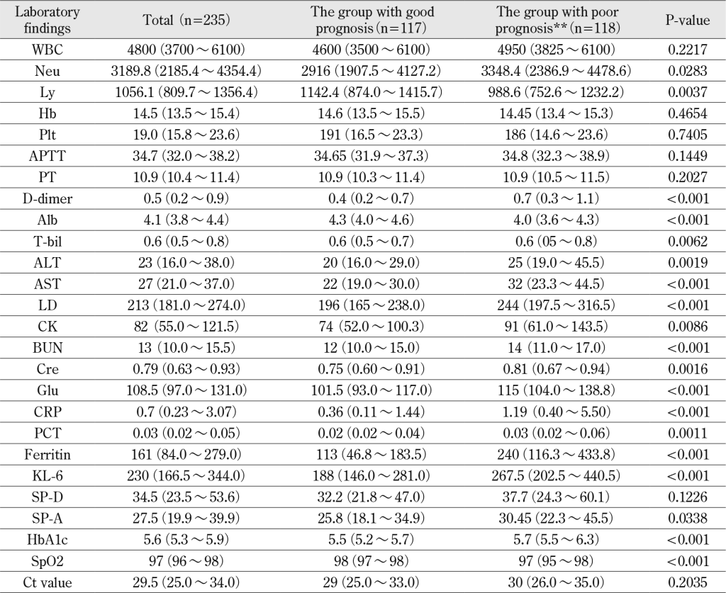
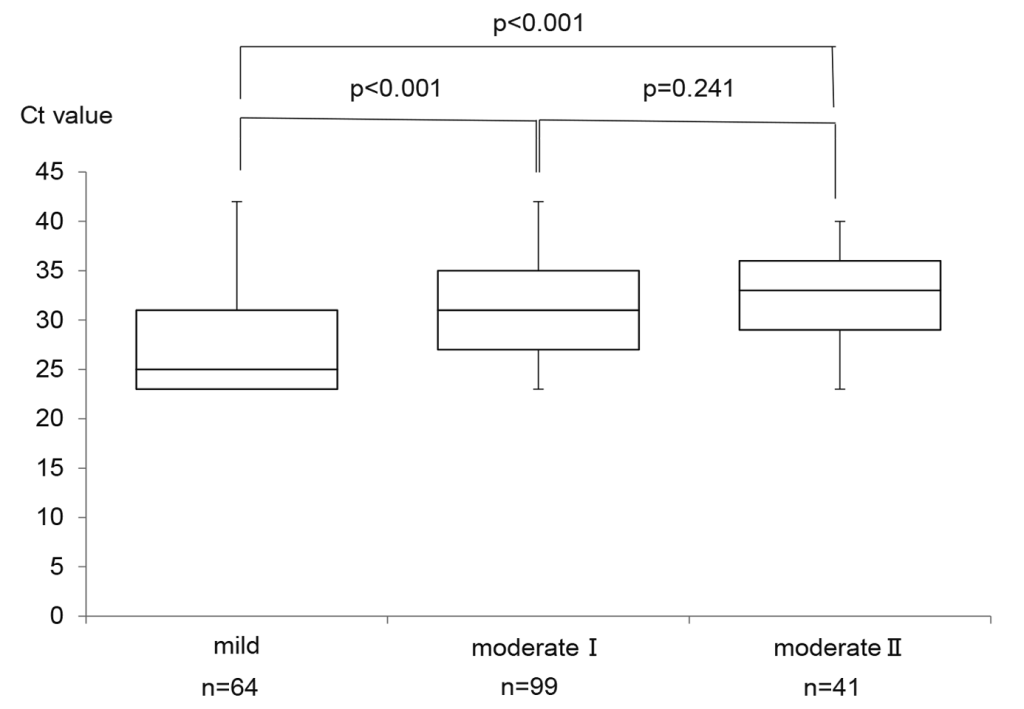
Laboratory findings at the initial visit in the poor prognosis group showed significant increases in neutrophil count (p=0.03), D-dimer (p<0.01), total bilirubin (p<0.01), ALT (p<0.01), AST (p<0.01), LD (p<0.01), CK (p<0.01), BUN (p<0.01), creatinine (p<0.01), glucose (p<0.01), CRP (p<0.01), procalcitonin (p<0.01), ferritin (p<0.01), KL-6 (p<0.01), SP-A (p=0.03), and HbA1c (p<0.01), and significant decreases in lymphocyte count (p<0.01), albumin (p<0.01) and SpO2(p<0.01). The Ct value did not show statistical difference between the good and poor prognosis groups. Furthermore, the relationship between the Ct value and severity was evaluated. The Ct showed higher values as the severity increased (Table 2). In particular, Ct values were not statistically different between patients with moderate I and moderate II, however were statistically higher in patients with moderate I and moderate II than in those with mild in severity (Fig.1).
4.Factors related to poor prognosis in patients with COVID-19
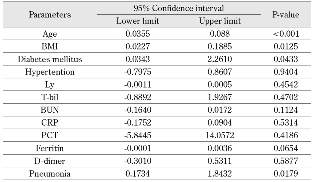
Multivariate analysis of prognostic factors considering the results of univariate analysis and previously reported prognostic factors showed that age (p<0.01), BMI (p=0.01), diabetes mellitus (p=0.04), and concurrent pneumonia (p=0.02) were independent prognostic factors (Table 3).
Cohort 2
Background and prognostic factors of patients with COVID-19
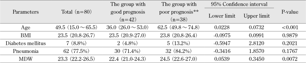
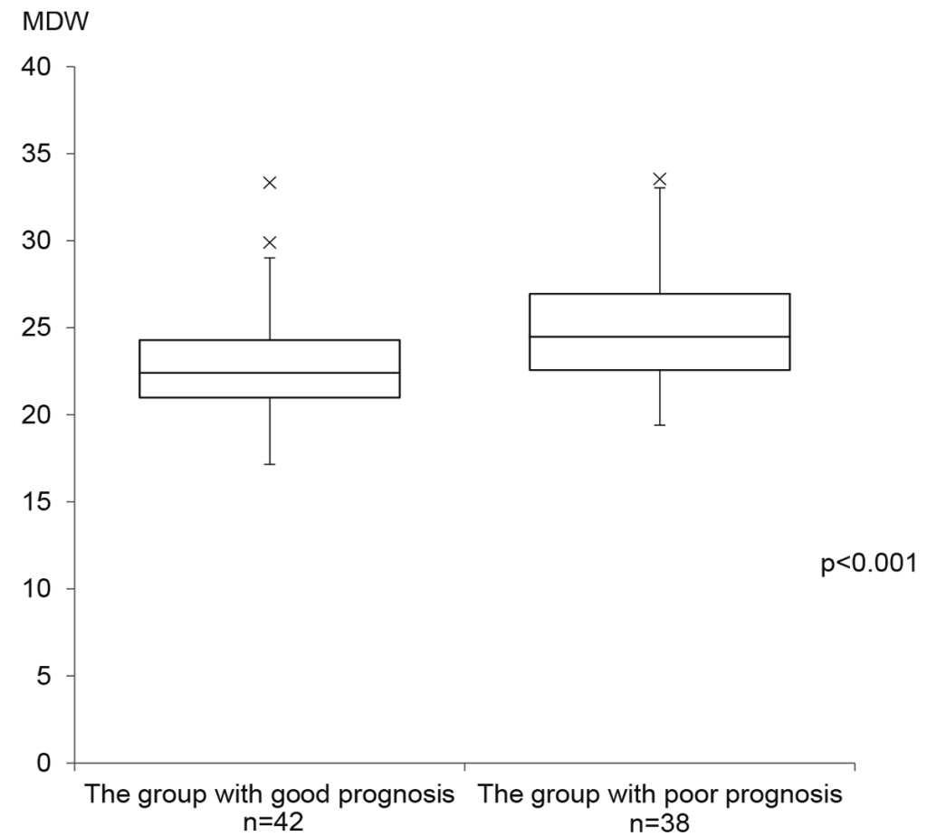
Since age, BMI, diabetes mellitus, and concurrent pneumonia were found to be independent prognostic factors of COVID-19 in Cohort 1, we also evaluated MDW as a prognostic factor in addition to age, BMI, diabetes mellitus, and concurrent pneumonia. The results showed significantly higher values for age and MDW (Table 4, Fig. 2).
IV.Discussion
Prognostic factors and markers of aggravation of COVID-19 have been investigated in many studies 24)- 34). However, there have been no reports on studies of the presence/absence of pneumonia based on chest CT images or studies including viral load at the initial visit to the hospital. There are not many studies of MDW related to COVID-19.
Prognosis should essentially be evaluated based on survival/death. However, since no deaths were observed in this study, poor prognosis was defined as patients who were hospitalized for 14 days or longer or who were transferred to other facilities due to exacerbation of symptoms after hospitalization, while good prognosis was defined as patients who were hospitalized for shorter than 14 days. As a result of multivariate analysis, age, BMI, diabetes mellitus, and concurrent pneumonia are found as independent prognostic factors in our study.
Advanced age was consistently found to be a prognostic factor in most of the previously reported studies in COVID-19 24), 28), 30), 33). High BMI 25), 28) and diabetes mellitus 24) are also often reported to be poor prognostic factors.
Malik P et al. investigated biomarkers of COVID-19 aggravation and reported that lymphopenia, thrombocytopenia, elevated D-dimer, elevated CRP, elevated procalcitonin, elevated AST, elevated ALT, elevated creatinine, and elevated LD were significantly associated with mechanical ventilation and death 34). This report is almost consistent with the results of univariate analysis in our study. Therefore, it was considered that the definitions of the poor and the good prognosis groups might be reasonable in view of the analysis of prognostic factors.
The severity of concurrent pneumonia is assessed as moderate or higher according to the 6.2th version of the Clinical Practice Guidelines 2), however it is unknown whether concurrent pneumonia on admission is related to subsequent aggravation. In some cases, pneumonia will be observed after hospitalization and become severe. COVID-19 pneumonia is considered to appear a few days after the onset of COVID-19. However, since the median time from the onset of COVID-19 infection to hospitalization in the patients investigated in this study was 5 days, the patients in whom COVID-19 infection became severe may have had pneumonia at the time of hospital admission.
According to a systematic review by Shah VP. et al., hospitalized patients with lower Ct value (i.e., higher levels of viral RNA) have higher disease severity and mortality, however it is also noted that the results of this analysis should be interpreted with caution, given the limitations and lack of assay standardization 35).
Our results showed that the viral load (Ct value) on admission was not associated with prognosis, and was statistically lower in patients with higher severity. It has already been reported that the viral load is the highest immediately after onset, and that there becomes no isolation of the virus from the 7th day after onset 36). The results of the Ct value in this study may suggest that the immune response plays a stronger role in the aggravation of COVID-19 than the virus itself, and that COVID-19 pneumonia is caused by the immune response. The Ct value does not only provide useful information for infection control since the viral concentration in specimens and the estimation of time of onset, but also might be used actively for the selection of antiviral and anti-inflammatory drugs.
Although many studies have been conducted on markers of aggravation, it is desirable to predict the prognosis of COVID-19 easily within the scope of routine clinical examinations.
MDW generated by DxH 900 which was evaluated in this study, is a new cytometric parameter that reflects monocyte changes in cell volume caused by the activation of monocytes. The result is obtained in around 1 minute with routine CBC and WBC differential testing without the need for ordering additional tests. Therefore, MDW values can be easily confirmed as a routine clinical examination. MDW may be considered a promising screening test parameter for COVID-19, if it’s prognostic prediction for COVID-19 is superior.
Originally, monocytes play an important role in the innate immune system against infection, and are believed to be involved in phagocytosis, antigen presentation, cytokine production, and activation of acquired immune system. Additionally, activation of monocytes is considered to result in the expression of various functions and the diversity of morphology 37)-42). Similarly, neutrophil volume and distribution width are changed, however, Crouser et al.43)-46) evaluated MDW in patients in the emergency department and reported that MDW was superior in detecting sepsis patients and effective as the initial biomarker to aid in diagnosis and severity assessment of infection.
It is also reported that MDW may be effective as a diagnostic aid and a severity assessment biomarker for COVID-19. Ognibene et al.47) reported an observational study analyzing 147 patients suspected of having COVID-19 who visited the emergency room. The mean MDW was 27.3±4.9 in the SARS-CoV-2 -positive patients (n=41) and 20.3±3.3 (p<0.005) in the SARS-CoV-2 -negative patients (n=106). In addition, when the MDW of 23 patients admitted to the ICU and 18 patients not admitted to the ICU were compared among SARS-CoV-2 positive patients, the mean values were 28.8±5.3 and 25.4±3.6 (P<0.05), respectively, and was significantly higher in the patients admitted to the ICU, suggesting that MDW is useful for the diagnosis and severity evaluation of COVID-19. However, it was not evaluated as a biomarker of aggravation in that study. Investigations as biomarkers of severe disease have been reported by Riva G, et al.48) and Frugoli A, et al.49)
Riva G, et al. studied 71 good prognosis cases (81.6%) and 16 poor prognosis cases (deaths) (18.4%) and reported that the last MDW value detected during the follow-up after diagnosis in each patient (n=87) was significantly higher in the poor prognosis group with a median value of 26.1 than in the good prognosis group. Frugeli et al. also studied 284 good prognosis cases (85.54%) and 47 poor prognosis cases (deaths) (14.16%) and reported that the median value at the first visit in the poor prognosis group was 24.9, significantly higher than in the good prognosis group. In our study, MDW was evaluated as a biomarker of aggravation, and MDW was significantly higher in the poor prognosis group. Riva G, et al. showed higher values than our results because they looked at the last value of MDW detected during the follow-up after diagnosis, but were in close agreement with the results of Frugeli et al.
Frugoli A, et al. also showed the usefulness of MDW as a predictive marker of sepsis in COVID-19 patients. In the present study, PCT was significantly higher in the poor prognosis group in cohort 1, suggesting that poor prognosis cases may have concurrent sepsis. This point may need further investigation with a larger number of cases.
These results suggest that MDW may be used as a biomarker of aggravation at the time of hospital admission.
This study has some limitations. First, prognosis should essentially be evaluated based on survival/death. However, since no deaths were observed in this study, poor prognosis was defined as patients who were hospitalized for 14 days or longer or who were transferred to other facilities due to exacerbation of symptoms after hospitalization, while good prognosis was defined as patients who were hospitalized for shorter than 14 days. Second, in this study, the prognostic factors were analyzed from July 2020, however since DxH 900 was installed in April 2021, the number of patients in whom MDW was analyzed was small and it was hard to examine in the same cohort. Third, the study was retrospective in design, used data obtained from a single center study. Finally, it did not include severe patients.
V. Conclusions
Viral load (Ct value) at the initial visit to the hospital may be not related to prognosis. However, it does not only provide useful information for infection control since viral concentration in the specimens and the time of onset may be estimated, but also might be used actively for the selection of antiviral and anti-inflammatory drugs.
MDW is a test that can be performed quickly and easily in peripheral blood testing, which is collected in a routine clinical examination and it is not necessary to use nasopharyngeal swab samples, etc. MDW is expected to be widely used clinically as a supplementary test to predict the prognosis of COVID-19 in various medical institutions such as small and medium-sized hospitals and clinics.
Declarations
Ethics approval and consent to participate
This study was conducted after being approved by the ethics committee of National Hospital Organization Omuta National Hospital (Approval No.: 3-19).
Competing interests
The authors declare that they have no conflicts of interest and no competing interests.
Funding
No funding was received for conducting this study.
Authors’ contributions
K. W. assisted with study design and interpretation of the data, had full access to the study data, assumes responsibility for the integrity of the data and the accuracy of the analysis, and drafted the manuscript. Z. N., K. K., M. Y., S. K., A. K., J. O., S. M., N. N., M. K., S.A. and H. Y. assisted with study design and interpretation of the data and edited the initial draft of the manuscript. I. F. and R. K. contributed to data collection and management. H. K. contributed to the categorization of patients with COVID-19.
Acknowledgements
None.
References
- Li Q, Guan X, Wu P, et al. Early Transmission Dynamics in Wuhan, China, of Novel Coronavirus-Infected Pneumonia. N Engl J Med 2020; 382(13): 1199-207.PubMed
- Novel coronavirus infection (COVID-19) medical practice guidelines version 6.2. 2022. https://www.mhlw.go.jp/content/000888608.pdf. Accessed 27 Jan 2022.
- Wakamatsu K, Katsuki K, Ikeda T, et al. Study of threshold cycle (Ct Value) in SARS-CoV-2 RT-PCR test with smart gene. Jpn J Med Pharm Sci. 2021; 78: 643-51.
- Crouser ED, Parrillo JE, Seymour C, et al. Improved early detection of sepsis in the ED with a novel monocyte distribution width biomarker. Chest 2017; 152(3): 518-26.PubMed
- Crouser ED, Parrillo JE, Seymour CW, et al. Monocyte distribution width: a novel indicator of sepsis-2 and sepsis-3 in high-risk emergency department patients. Crit Care Med 2019; 47(8): 1018-25.PubMed
- Crouser ED, Parrillo JE, Martin GS, et al. Monocyte distribution width enhances early sepsis detection in the emergency department beyond SIRS and qSOFA. J Intensive Care 2020; 8: 33.PubMed
- Woo A, Oh DK, Park CJ, et al. Monocyte distribution width compared with C-reactive protein and procalcitonin for early sepsis detection in the emergency department. PLoS One 2021; 16(4): e0250101.PubMed
- Hausfater P, Boter NR, Indiano CM, et al. Monocyte distribution width (MDW) performance as an early sepsis indicator in the emergency department: comparison with CRP and procalcitonin in a multicenter international European prospective study. Crit Care 2021; 25(1): 227.PubMed
- Agnello L, Bivona G, Vidali M, et al. Monocyte distribution width (MDW) as a screening tool for sepsis in the emergency department. Clin Chem Lab Med 2020; 58(11): 1951-7.PubMed
- Agnello L, Sasso BL, Giglio RV, et al. Monocyte distribution width as a biomarker of sepsis in the intensive care unit: a pilot study. Ann Clin Biochem 2021; 58(1): 70-3.PubMed
- Piva E, Zuin J, Pelloso M, et al. Monocyte distribution width (MDW) parameter as a sepsis indicator in intensive care units. Clin Chem Lab Med 2021; 59(7): 1307-14.PubMed
- Guo C, Li B, Ma H, et al. Single-cell analysis of two severe COVID-19 patients reveals a monocyte-associated and tocilizumab-responding cytokine storm. Nat Commun 2020; 11(1): 3924.PubMed
- Chousterman BG, Swirski FK, Weber GF. Cytokine storm and sepsis disease pathogenesis. Semin Immunopathol. 2017; 39(5): 517-28.PubMed
- Mukherjee R, Barman PK, Thatoi PK, et al. Non-classical monocytes display inflammatory features: validation in sepsis and systemic lupus erythematous. Sci Rep. 2015; 5: 13886.PubMed
- Sprangers S, De Vries TJ, Everts V. Monocyte heterogeneity: consequences for monocyte-derived immune cells. J Immunol Res 2016; 2016: 1475435.PubMed
- Cormican S, Griffin MD. Human monocyte subset distinctions and function: insights from gene expression analysis. Front Immunol 2020; 11: 1070.PubMed
- Kaur G, Sandeep F, Olayinka O, et al. Morphologic changes in circulating blood cells of COVID-19 patients. Cureus 2021; 13(2): e13416.PubMed
- Mehta P, McAuley DF, Brown M, et al. COVID-19: consider cytokine storm syndromes and immunosuppression. Lancet 2020; 395(10229): 1033-4.PubMed
- Huang C, Wang Y, Li X, et al. Clinical features of patients infected with 2019 novel coronavirus in Wuhan, China. Lancet 2020; 395(10223): 497-506.PubMed
- Wakamatsu K, Nagasawa Z, Katsuki K, et al. Retrospective study on the efficacy of monocyte distribution width (MDW) as a screening test for COVID-19. European Journal of Medical Research 2023; 28(1): 136. doi: 10.1186/s40001-023-01086-7.PubMed
- Ognibene A, Lorubbio M, Magliocca P, et al. Elevated monocyte distribution width in COVID-19 patients: the contribution of the novel sepsis indicator. Clin Chim Acta 2020; 509: 22-4.PubMed
- Lin HA, Lin SF, Chang HW, et al. Clinical impact of monocyte distribution width and neutrophil-to-lymphocyte ratio for distinguishing COVID-19 and influenza from other upper respiratory tract infections: a pilot study. PLoS One 2020; 15(11): e0241262.PubMed
- Riva G, Castellano S, Nasillo V, et al. Monocyte distribution width (MDW) as novel inflammatory marker with prognostic significance in COVID-19 patients. Sci Rep 2021; 11(1): 12716.PubMed
- Terada M, Ohtsu H, Saito S, et al. Risk factors for severity on admission and the disease progression during hospitalization in a large cohort of patients with COVID-19 in Japan. BMJ Open 2021; 11(6): e047007. doi:10.1136/bmjopen-2020-047007.PubMed
- de Siqueira JVV, Almeida LG, Zica BO, et al. Impact of obesity on hospitalizations and mortality, due to COVID-19: A systematic review. Obes Res Clin Pract 2020; 14(5): 398-403. doi:10.1016/j.orcp.2020.07.005.PubMed
- Pranata R, Lim MA, Huang I, et al. Hypertension is associated with increased mortality and severity of disease in COVID-19 pneumonia: A systematic review, meta-analysis and meta-regression. J Renin Angiotensin Aldosterone Syst 2020; 21(2):1470320320926899. doi:10.1177/1470320320926899.PubMed
- Lippi G, Wong J, Henry BM. Hypertension in patients with coronavirus disease 2019 (COVID-19): a pooled analysis. Pol Arch Intern Med 2020; 130(4): 304-9. doi:10.20452/pamw.15272.PubMed
- Fried MW, Crawford JM, Mospan AR, et al. Patient Characteristics and Outcomes of 11,721 Patients with Coronavirus Disease 2019 (COVID-19) Hospitalized Across the United States. Clin Infect Dis 2021; 72(10): e558-65. doi:10.1093/cid/ciaa1268.PubMed
- Wang B, Li R, Lu Z, et al. Does comorbidity increase the risk of patients with COVID-19: evidence from meta-analysis. Aging (Albany NY) 2020; 12(7): 6049-57. doi:10.18632/aging.103000.PubMed
- Fang X, Li S, Yu H, et al. Epidemiological, comorbidity factors with severity and prognosis of COVID-19: a systematic review and meta-analysis. Aging (Albany NY) 2020; 12(13): 12493-503. doi:10.18632/aging.103579.PubMed
- Zhou Y, He Y, Yang H, et al. Development and validation a nomogram for predicting the risk of severe COVID-19: A multi-center study in Sichuan, China. PLoS One 2020; 15(5): e0233328. doi:10.1371/journal.pone.0233328.PubMed
- Patel U, Malik P, Usman MS, et al. Age-Adjusted Risk Factors Associated with Mortality and Mechanical Ventilation Utilization Amongst COVID-19 Hospitalizations-a Systematic Review and Meta-Analysis. SN Compr Clin Med 2020; 2(10): 1740-9. doi:10.1007/s42399-020-00476-w.PubMed
- Harrison SL, Fazio-Eynullayeva E, Lane DA, et al. Comorbidities associated with mortality in 31,461 adults with COVID-19 in the United States: A federated electronic medical record analysis. PLoS Med 2020; 17(9): e1003321. doi:10.1371/journal.pmed.1003321.PubMed
- Malik P, Patel U, Mehta D, et al. Biomarkers and outcomes of COVID-19 hospitalisations: systematic review and meta analysis. BMJ Evi Based Med 2021; 26(3): 107-8. doi: 10.1136/bmjebm-2020-111536. Epub 2020 Sep 15.PubMed
- Shah VP, Farah WH, Hill JC, et. al. Association Between SARS-CoV-2 Cycle Threshold Values and Clinical Outcomes in Patients With COVID-19: A Systematic Review and Meta-analysis. Open Forum Infect Dis 2021; 8(9): ofab453.PubMed
- Sethuraman N, Jeremiah SS, Ryo A. Interpreting Diagnostic Tests for SARS-CoV-2. JAMA 2020; 323(22): 2249-51.PubMed
- Guo C, Li B, Ma H, et al. Single-cell analysis of two severe COVID-19 patients reveals a monocyte-associated and tocilizumab-responding cytokine storm. Nat Commun 2020; 11(1): 3924.PubMed
- Chousterman BG, Swirski FK, Weber GF. Cytokine storm and sepsis disease pathogenesis. Semin Immunopathol 2017; 39(5): 517-28.PubMed
- Mukherjee R, Barman PK, Thatoi PK, et al. Non-classical monocytes display inflammatory features: validation in sepsis and systemic lupus erythematous. Sci Rep 2015; 5: 13886.PubMed
- Sprangers S, De Vries TJ, Everts V. Monocyte heterogeneity: consequences for monocyte-derived immune cells. J Immunol Res 2016; 2016: 1475435.PubMed
- Cormican S, Griffin MD. Human monocyte subset distinctions and function: insights from gene expression analysis. Front Immunol 2020; 11: 1070.PubMed
- Kaur G, Sandeep F, Olayinka O, et al. Morphologic changes in circulating blood cells of COVID-19 patients. Cureus 2021; 13(2): e13416.PubMed
- Crouser ED, Parrillo JE, Seymour C, et al. Improved early detection of sepsis in the ED with a novel monocyte distribution width biomarker. Chest 2017; 152(3): 518-26.PubMed
- Crouser ED, Parrillo JE, Seymour C, et al. Improved early detection of sepsis in the ED with a novel monocyte distribution width biomarker. Chest 2017; 152(3): 518-26.
- Crouser ED, Parrillo JE, Seymour CW, et al. Monocyte distribution width: a novel indicator of sepsis-2 and sepsis-3 in high-risk emergency department patients. Crit Care Med 2019; 47(8): 1018-25.PubMed
- Crouser ED, Parrillo JE, Martin GS, et al. Monocyte distribution width enhances early sepsis detection in the emergency department beyond SIRS and qSOFA. J Intensive Care 2020; 8: 33.PubMed
- Ognibene A, Lorubbio M, Magliocca P, et al. Elevated monocyte distribution width in COVID-19 patients: the contribution of the novel sepsis indicator. Clin Chim Acta. 2020; 509: 22–4.PubMed
- Riva G, Castellano S, Nasillo V, et al. Monocyte Distribution Width (MDW) as novel inflammatory marker with prognostic significance in COVID-19 patients. Sci Rep 2021; 11(1): 12716. doi: 10.1038/s41598-021-92236-6PubMed
- Frugoli A, Ong J, Meyer B, et al. Monocyte Distribution Width Predicts Sepsis, Respiratory Failure, and Death in COVID-19. Cureus 2023; 15(12): e50525. doi:10.7759/cureus.50525.PubMed