
Marie Yamada1, Naotomo Yamada1, †Yasushi Kubota2,3, Emi Takahashi4, Mami Nakao1, Toru Miyazaki4, Shinichiro Sato4, Hideaki Nakamura1, Shinya Kimura2, †Eisaburo Sueoka5
Yamada M, Yamada N, Kubota Y, Takahashi E, Nakao M, Toru Miyazaki4, Shinichiro Sato4, Hideaki Nakamura1, Shinya Kimura2, †Eisaburo Sueoka5. A patient with partial E phenotype expressing alloanti-Eantibody after transfusion with platelet concentrates. Lab Med Int 2024; 3(3): 66-69. doi: 10.51041/lmi.3.3_66
Case Report
Lab Med Int 2024; 3(3): 66-69
†Correspondence: Department of Transfusion and Cell Therapy, Saitama Medical Center, Saitama Medical University, 1981 Kamoda, Kawagoe 350-8550, Japan. E-mail: kubotay”@”saitama-med.ac.jp.
Department of Clinical Laboratory Medicine, Faculty of Medicine, Saga University, 5-1-1 Nabeshima, Saga 849-8501, Japan.
E-mail: sueokae”@”cc.saga-u.ac.jp
Received November 27, 2023; accepted July 18, 2024
*1 Department of Transfusion Medicine, Saga University Hospital, 5-1-1 Nabeshima, Saga 849-8501, Japan.
*2 Division of Hematology, Respiratory Medicine and Oncology, Department of Internal Medicine, Faculty of Medicine, Saga University, 5-1-1 Nabeshima, Saga 849-8501, Japan.
*3 Department of Transfusion Medicine and Cell Therapy, Saitama Medical Center, Saitama Medical University, 1981 Kamoda, Kawagoe 350-8550, Japan.
*4 Laboratory section of Japanese Red Cross Hokkaido Block Blood Center, Sapporo, Japan.
*5 Department of Clinical Laboratory Medicine, Faculty of Medicine, Saga University, 5-1-1 Nabeshima, Saga 849-8501, Japan.
ABSTRACT
Partial E is a qualitative mutation of the E antigen that lacks one or more components of the E antigen, and several variants have been reported. We report a 72-year-old man having partial E(allele name: RHCE*cEFM)who expressed alloanti-E antibody after platelet concentrates transfusions. The patient was diagnosed with relapsed non-Hodgkin’s lymphoma and received salvage chemotherapy. One platelet transfusion was from a donor with the R1R2 phenotype when he received three rounds of transfusions of 10 units platelet concentrate from February to April 2009 for myelosuppression by chemotherapy. The patient, who had no history of transfusion prior to 2009, was sensitized by small amounts of allogeneic red blood cells contaminating platelet concentrates and developed alloanti-E antibodies. The anti-E antibody was detected in the patient’s serum, and Rh phenotype determination revealed relatively weak reactivity (2+) for a monoclonal anti-E reagent but negative for another one. According to these results, an E variant phenotype was suspected. Direct sequencing of exon 5 on the RHCE gene revealed mutations of C to G at nucleotide c.697 and A to G at nucleotide c.712 in the E allele, resulting in amino acid substitutions phenylalanine to alanine and methionine to valine, respectively. Combining this result with reactivity based on an antigen screening test for E antigen using monoclonal antibodies, we classified the patient’s RBC phenotype as partial E due to RHCE alterations. To our knowledge, this is the first case report of the RHCE rare variant patient with alloanti-E antibody produced by platelet concentrates transfusions.
〔Lab Med Int 2024; 3(3): 66-69〕
Key Words
Rh blood type, E antigen, Partial E blood type, RHD gene
I. Introduction
Partial E blood type is a rare E variant that lacks some of the Rh system antigens1)-5). There are at least 5 types of partial E in the genetic background. These include a single nucleotide exchange T to A at codon 500 in the RHCE gene, resulting in amino acid substitution of methionine to lysine at codon 167 (allele name: RHCE*cEKK); replacement of exons 1, 2, and 3 in the RHCE gene with the RHD gene, resulting in a hybrid RHD-CE gene (allele name: RHCE*cEKK); a rearrangement of exon 5 in the RHCE gene with the RHD gene; and amino acid substitutions glutamic acid to glutamine at codon 233 and methionine to valine at codon 238, resulting in the RHCE-D-CE variant hybrid gene (allele name: RHCE*cEKK); amino acid substitution arginine to threonine at codon 201, which locates in transmembrane domain 6, resulting in the RHCE-D-CE rearranged gene (allele name: RHCEcIV); and a one-base mutation G to A at codon 461 with amino acid substitution arginine to threonine at codon 154 in the RHCE gene (allele name: RHCE*cEKH)3),4).
Here we report the unique clinical course of a non-Hodgkin’s lymphoma (NHL) patient who expressed alloanti-E antibodies after platelet transfusion, and the results of genetic and serological analysis for RhE phenotype.
II. Case and clinical course
In 2009, a seventy-two-year-old man was admitted to our hospital because of recurrence of NHL. His blood type was A and RhD positive based on serological tests. His medical history showed no blood transfusions before 2009, and in 2006 he tested negative for irregular antibodies as determined by the low ionic strength solution-enhanced indirect anti-globulin test (LISS-IAT) and the polyethylene glycol-enhanced indirect anti-globulin test (PEG-IAT).
The patient received three rounds of transfusions of 10 units platelet concentrate between February and April 2009 due to myelosuppression caused by chemotherapy; after these transfusions, he also received red blood cell (RBC)concentrates because of severe anemia. Screening for irregular antibodies before RBC transfusion was conducted and returned a result of positive for anti-E antibody. Precise examination for Rh phenotypes showed positive agglutination of RBCs to the following monoclonal antibodies: anti-C (4+), anti-c (3+), anti-E (w+), and anti-e (3+), as well as weak activity for monoclonal anti-E reagent. Reactivity for human type A anti-E sera was positive (1+)by PEG-IAT, and no reactivity between the patient’s own RBC and patient serum with anti-E antibody was observed by PEG-IAT.
In 2013 the patient was readmitted to our hospital due to a second relapse of NHL, at which time he received salvage chemotherapy. Severe anemia (hemoglobin 5.4 g/dL) was observed so he was given RBC transfusions. Antibody screening before RBC transfusion showed no irregular antibodies. Rh phenotype determination revealed relatively weak activity (2+) for monoclonal anti-E reagent (Ortho Bioclone®) and negative activity for another monoclonal anti-E reagent (Gamma-clone®). According to these results, an E variant was suspected and we requested further, more precise, evaluation of RhE phenotype and genotyping by the Japanese Red Cross Hokkaido Block Blood Center.
III. Methods
- Serological tests
Twenty-three monoclonal anti-E antibodies, which are approved by the 4th International Workshop on Monoclonal Antibodies Against Human Red Blood Cells and Related Antigens, were used for evaluation of RhE phenotype. R1R2 RBC and rr RBC were used for positive and negative controls, respectively. Four types of unclassified partial E RBCs were also analyzed in these assays. - Genotyping
Genomic DNA was extracted from whole blood. Specific primers for exons 3, 4, and 5 of the RHCE gene were designed and genomes amplified by PCR were sequenced. Primer sets used for the RHCE gene are as follows: exon 3 sense 5’-ATCCTGGCTCTCCTTCTCA-3’, anti-sense 5’-CAAGTGATCTTCCCTCCTCAA-3’; exon 4 sense 5’-TGAACTTTCTCCAAGGACCAT-3’, anti-sense 5’-AATTTAGCAAACACTACTCAAAGAAG-3’; exon 5 sense 5’-GCAACAGAGCAAGAGTCCA-3’, anti-sense 5’-GTGACCACCCAGCATTCTT-3’. - Ethics
This study was approved by the Institutional Review Board of Saga University Hospital (2020-02-R-11).
IV. Results
Serological examinations
The patient’s RBCs showed no reactivity against 13 of the 23 monoclonal anti-E antibodies examined (Table 1). They showed reactive patterns that resembled one of the unclassified partial E RBCs against monoclonal anti-E antibodies (Table 1).
Genotyping
Exons 3 and 4 of the RHCE gene had no mutation. Heterozygous mutation peaks were observed at nucleotides c.676 (C to G), c.697 (C to G), and c.712 (A to G) in the E allele. Among them, the c.676C>G (p.Pro226Ala) mutation is responsible for the expression of e antigen, which was positive in the patient’s RBCs. In addition, the c.697C>G and c.712A>G sequences are identical with the sequences in exon 5 of the RHD gene. These results suggest that a partial region in exon 5 of the RHCE gene was substituted into a homologous region in exon 5 of the RHD gene, resulting in an RHCE-D-CE gene containing amino acid substitutions p.Gln233Glu and p.Met238Val, producing a partial E phenotype (allele name: RHCE*cEFM) (Fig. 1).
V. Discussion
In the literature, five types of partial E have been evaluated genetically3),4). In addition to the partial E phenotype, Ew (RH11) phenotype─a rare variant form of E antigen showing unique reactivity against anti-E antibodies─has been reported, and an individual carrying the Ew (RH11) produced alloanti-E antibodies6)-8). Our patient expressed alloanti-E antibody in his sera after being given platelet transfusions. The appearance of irregular antibodies to allo-RBCs after transfusions with platelet concentrates has been reported in several cases9),10). Our patient received platelets from a donor who was carrying the R1R2 phenotype, suggesting that the patient was sensitized by small amounts of allo-RBCs contaminating the platelet concentrates. The subtype of detected anti-E antibody in the patient could not be determined conclusively, but was probably IgG because the antibody was negative for assay with saline and positive (1+) for PEG-IAT. Furthermore, no reactivity between the patient’s own RBCs and patient serum with anti-E antibodies was observed by PEG-IAT, suggesting that the anti-E antibody was induced by allo-RBCs. In the present case, since anti-E has been detected, the appropriate response is to transfuse E antigen-negative red blood cell products.
In summary, we report a rare variant of Rh E antigen, partial E (allele name: RHCE*cEFM). This is the first case report of partial E (RHCE*cEFM) genetically confirmed in a Japanese patient with alloanti-E antibody. Our case possesses alloanti-E antibodies to the deficient part of the E antigen, possibly due to exposure from allo-RBCs expressing the E antigen in platelet concentrates. Testing with multiple monoclonal anti-E reagents or RHCE gene analysis is important if the reaction with the monoclonal anti-E reagent is weak because of the possibility of partial E.
Table 1 Reactive patterns of the patient’s RBCs against various monoclonal anti-E antibodies
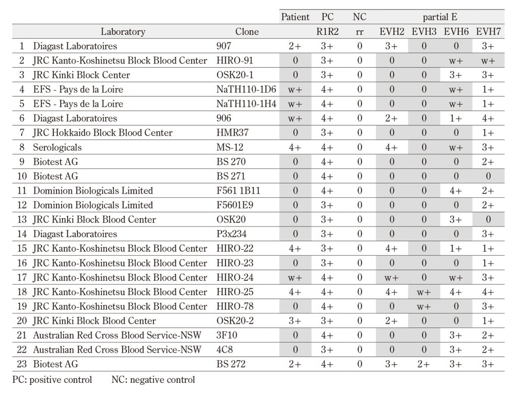
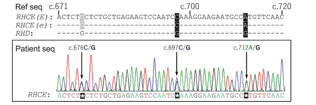
Figure 1 Three heterozygous peaks were observed in the patient RHCE exon5. The upper panel indicates a part of
reference sequence of RHCE( NM_020485.5) and RHD( NM_016124.4) exon5. Gray background at c.676 in
RHCE is responsible for the expression of e or E antigen. Black background at c.697 and c.712 means different
nucleotide between RHCE and RHD. Hyphen in the RHD sequence and the RHCE (e) sequence indicates
homology with the RHCE (E) sequence. We analyzed the patient sequence of RHCE exon3, 4 and 5 by using
genomic DNA from whole blood and specific primers. The lower panel indicates a part of the direct sequence
data of RHCE( from c.671 to c.720) of the patient. White asterisks in black background( c.676, c.697 and c.712)
represents the heterozygous peak of patient RHCE exon5.
Acknowledgments
This work was supported in part by the following Grants-in Aid for Cancer Research: Special Cancer Research, from the Ministry of Education, Science, Sports and Culture, Japan, and for the Research on Hematopoietic Stem Cell Preservation Study from Saga Prefecture, Japan, and Saitama Medical University Research Grant (No. 23-B-1-04).
Authorship
Contributions: M.Y., N.Y., and Y.K. performed the analyses, interpreted the data and wrote the paper. E.T., T.M., and S.S. performed serological tests and genotyping for RhE phenotype. M.N., H.N., and S.K. interpreted the data. E.S. designed the research, interpreted the data, and wrote the paper. In addition, all authors provided their final approval of the manuscript.
Conflicts of Interest
The authors declare that they have no conflict of interest.
References
- Avent ND, Reid ME. The Rh blood group system: a review. Blood 2000; 95(2): 375-87.PubMed
- Avent ND, Finning KM, Liu W, et al. Molecular biology of partial D phenotypes. Transfus Clin Biol 1996; 3(6): 511-6. doi: 10.1016/s1246-7820(96)80073-5.PubMed
- Noizat-Pirenne F, Mouro I, Gane P, et al. Heterogeneity of blood group RhE variants revealed by serological analysis and molecular alteration of the RHCE gene and transcript. Br J Haematol 1998; 103(2): 429-36. doi: 10.1046/j.1365-2141.1998.01004.x.PubMed
- Kashiwase K, Ishikawa Y, Hyodo H, et al. E variants found in Japanese and c antigenicity alteration without substitution in the second extracellular loop. Transfusion 2001; 41(11): 1408-12. doi: 10.1046/j.1537-2995.2001.41111408.x.PubMed
- Chérif-Zahar B, Raynal V, D’Ambrosio AM, et al. Molecular analysis of the structure and expression of the RH locus in individuals with D–, Dc-, and DCw- gene complexes. Blood 1994; 84(12): 4354-60.PubMed
- Strobel E, Noizat-Pirenne F, Hofmann S, et al. The molecular basis of the Rhesus antigen Ew. Transfusion 2004; 44(3): 407-9. doi: 10.1111/j.1537-2995.2004.00655.x.PubMed
- Lubenko A, Burslem SJ, Fairclough LM, et al. A new qualitative variant of the RhE antigen revealed by heterogeneity among anti-E sera. Vox Sang 1991; 60(4): 235-40. doi: 10.1111/j.1423-0410.1991.tb00912.x.PubMed
- Henke J, Kasulke D. The first example of the Rh antigen Ew in Western Europe. Vox Sang 1976; 30(4): 305-7. doi: 10.1111/j.1423-0410.1976.tb02832.x.PubMed
- Bartley AN, Carpenter JB, Berg MP. D+ Platelet trans‑fusion in D-patients: cause for concern? Immunohematology 2009; 25(1): 5-8.PubMed
- Kitazawa J, Nollet K, Morioka H. Non‑D Rh anti‑bodies appearing after apheresis platelet transfusion: stimulation by red cells or microparticles? Vox Sang 2011; 100(4): 395-400. doi: 10.1111/j.1423-0410.2010.01435.x.PubMed