Satoshi Fujimura*1,2, †Yumiko Higuchi*1,3, Atsushi Izawa*1,4,5, Tomoki Ichikawa*1, Yoko Usami*2, Makoto Yamaura*1, Tsukasa Higuchi*6,7, Fumiko Terasawa*8, Nobuo Okumura*1,3
Cite
Fujimura S, Higuchi Y, Izawa A, Ichikawa T, Usami Y, Yamaura M, Higuchi T, Terasawa F, Okumura N. Evaluation of serum citrullinated fibrinogen in patients with acute aortic dissection. Lab Med Int 2024; 3(2): 50-58. doi: 10.51041/lmi.3.2_50
Original
Lab Med Int 2024; 3(2): 50-58
†Correspondence: Department of Health and Medical Sciences,Graduate School of Health Sciences Shinshu University 3-1-1 Asahi, Matsumoto 390-8621, Japan.
E-mail: sasa0922″@”shinshu-u.ac.jp
Received May 26, 2023; accepted March 25, 2024
*1 Department of Health and Medical Sciences, Graduate School of Medicine, Shinshu University, Matsumoto, Japan
*2 Department of Laboratory Medicine, Shinshu University Hospital, Matsumoto, Japan
*3 Department of Biomedical Laboratory Sciences, Shinshu University School of Health Sciences, Matsumoto, Japan
*4 Department of Nursing, Shinshu University School of Medicine, Matsumoto, Japan
*5 Department of Cardiovascular Medicine, Shinshu University School of Health Sciences, Matsumoto, Japan
*6 Department of General Pediatrics, Nagano Children’s Hospital, Azumino, Japan
*7 Life Science Research Center, Nagano Children’s Hospital, Azumino, Japan
*8 Faculty of Health and Medical Sciences, Department of Medical Technology and Clinical Engineering, Hokuriku University, Kanazawa, Japan
ABSTRACT
Background: Acute aortic dissection (AAD) is a life-threatening cardiovascular disease that requires rapid intervention. The present study assessed the serum citrullinated fibrinogen (C-Fbg) levels in patients with AAD and investigated their clinical utility.
Materials and methods: Serum C-Fbg concentrations were measured at the first blood sample of AAD patients (n = 34) and compared with those of healthy controls (HCs; n = 23) and patients with ischemic heart disease (IHD; n = 10). We also assessed the correlations between C-Fbg levels and inflammatory and coagulation markers in patients with early-phase AAD as well as the time course of these markers, including C-Fbg.
Results: Serum C-Fbg levels were significantly higher in AAD patients than in HCs (p<0.001) and IHD patients (p<0.01), and serum C-Fbg levels correlated with D-dimer but not with other markers. Receiver operating characteristic (ROC) curve analysis revealed that C-Fbg concentrations were effective in differentiating AAD from IHD. In contrast to the decrease in D-dimer levels, C-Fbg concentrations remained high and relatively unchanged in early-phase AAD.
Conclusion: Serum C-Fbg levels were elevated in patients with AAD, and could differentiate AAD from IHD. Although further studies are needed, C-Fbg may be an adjunctive tool in the differential diagnosis of AAD.
〔Lab Med Int 2024; 3(2): 50-58〕
Key Words
Citrullinated fibrinogen, Acute aortic dissection, Ischemic heart disease, D-dimer
I.Introduction
Acute aortic dissection (AAD) is a life-threatening cardiovascular disease associated with high mortality 1), and early diagnosis is required to improve its prognosis. Although there are still uncertainties concerning the mechanisms underlying the onset of aortic dissection 2), many studies have demonstrated that a certain level of inflammation involving neutrophil or macrophage infiltration into aortic tissue is associated with the formation of aortic dissection 1) 3)-8).
The diagnosis of AAD is primarily based on symptoms and imaging findings. Blood tests play a role in the acute setting and can enhance the differential diagnosis of AAD from other serious illnesses. Many biochemical assays have been developed to detect AAD in such patients 9)10), including evaluations of smooth muscle myosin 11), matrix metalloproteinase-9 12), elastin degradation products 13), calponin 14), transforming growth factor-beta15) and D-dimer 16) 17). Among them, serum D-dimer concentration is currently the only biomarker for distinguishing AAD from ischemic heart disease (IHD) in the clinical settings 18) 19). Therefore, other rapid and accurate biomarkers are needed to enhance the diagnosis of AAD in the acute phase.
Protein citrullination involves the posttranslational modification of arginine to citrulline via the calcium-dependent enzyme peptidylarginine deiminase (PAD) 20)21). The 5 PAD isozymes (PAD 1, 2, 3, 4, and 6) are tissue-specifically distributed in human body and citrullinate proteins in a substrate-specific manner 22). Among them, PAD2 and PAD4 are expressed in blood cells 23).
Citrullinated proteins have been investigated as antigens for autoimmune antibodies in rheumatoid arthritis 20) 21). Among them, citrullinated fibrinogen (C-Fbg) is a well-known candidate autoantigen abundant in the joints of patients with rheumatoid arthritis, where a complicated inflammatory response occurs24). However, little is known about the physiological or pathological role of C-Fbg in other diseases.
We previously showed that C-Fbg levels were increased in patients with bacteremia, which was correlated with neutrophilia25). As neutrophilia is detected soon after AAD occurs 26), we hypothesized that Fbg might be citrullinated by PAD derived from increased neutrophils in or around the aortic wall in patients with AAD, causing an increase in C-Fbg levels.
In the present study, we assessed the specificity and potential clinical use of C-Fbg as an acute-phase marker for the early diagnosis of patients with suspected AAD.
II.Materials and methods
Subjects
Patients enrolled in this study were admitted to the Advanced Emergency and Critical Care Center of Shinshu University Hospital from November 2017 to July 2018 and diagnosed with AAD (Stanford type A or B) and IHD (7 cases of acute myocardial infarction, 2 cases of angina, and 1 case of acute coronary syndrome). Patients diagnosed with an infectious disease or active autoimmune disease, such as rheumatoid arthritis or systemic lupus erythematosus, were excluded. In this study, 1–6 serum samples were obtained from 34 patients with AAD (n = 118) and 10 patients with IHD (n = 10) independent of treatment strategy. For healthy controls (HCs; n = 23), blood samples were obtained from healthy volunteers after informed consent was obtained. All serum samples were stored at -30℃ until analysis. Clinical laboratory data, including concentrations of C-reactive protein (CRP), Fbg, and D-dimer, complete blood count, and differential white blood cell counts, were collected from the patients’ medical records. The reference intervals of values at Shinshu University Hospital were used. The participants’ detailed information is provided in Table 1.
This study was conducted in accordance with The Code of Ethics of the World Medical Association (Declaration of Helsinki) and approved by the Ethical Review Board of Shinshu University School of Medicine (no. 3928).
Sandwich enzyme-linked immunosorbent assay for C-Fbg
C-Fbg was quantified using a sandwich enzyme-linked immunosorbent assay method that we previously established. Briefly, the generated anti-C-Fbg antibody (clone F) was used as a precoating antibody, while biotin-conjugated anti-Fbg polyclonal antibody (Ab6666; Abcam, Cambridge, UK) was used as the secondary antibody 25).
The synthesis, purification, and citrullination of recombinant fibrinogen used as a standard were described previously 27)-29).
Statistical analyses
All statistical analyses were performed using EZR software (Saitama Medical Center, Jichi Medical University, Saitama, Japan), a graphical user interface for R software (The R Foundation for Statistical Computing, Vienna, Austria). The Mann–Whitney U test was used to compare inflammatory and coagulation markers between patients with AAD and those with IHD. In addition, a receiver operating characteristic (ROC) curve analysis was performed for C-Fbg concentration in patients with AAD and IHD. The optimal cut-off value was determined using Youden’s index. The Kruskal–Wallis test with the Steel–Dwass test was also used to compare C-Fbg concentrations between patients with AAD and those with IHD in the first submission sample and the HCs, as were chronological changes in C-Fbg and other markers. Moreover, the time-course transition of C-Fbg level and neutrophil count was evaluated using the same statistical analysis. Spearman’s rank correlation test was used to determine the correlation between serum C-Fbg level and other clinical factors. P values < 0.05 were considered statistically significant.
Table 1 Clinical characteristics of the patients and healthy controls
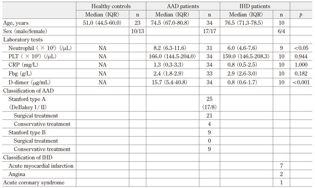
AAD: acute aortic dissection, CRP: C-reactive protein, Fbg: fibrinogen. IHD: IHD: ischemic heart disease, IQR: interquartile range, NA: not analyzed, PLT: platelet count.
III.Results
Initial serum C-Fbg concentrations of patients with AAD versus IHD
First, we evaluated the serum C-Fbg concentration in the first submitted sample of patients with AAD and compared them to those of patients with IHD and HCs. As illustrated in Figure 1A, the serum C-Fbg level in the first sample of AAD patients (median [range], 153.8 [101.2–218.6] ng/mL) was significantly higher than those of IHD patients (83.1 [74.0–92.9] ng/mL; p < 0.01) and HCs (40.5 [31.5–50.6] ng/mL; p < 0.001). A significant difference was also observed in the C-Fbg concentrations between the IHD patients and HCs. There were no significant differences in C-Fbg concentration between the cases of Stanford type A and B (Figure 1B) or DeBakey I and II (data not shown) in the AAD group.
The ROC analysis of C-Fbg concentrations in AAD and IHD patients in the first samples revealed an area under the curve (AUC) of 0.821 (95% confidence interval, 0.667–0.974) with a cut-off value of 94.1 ng/mL, sensitivity of 80.0%, and specificity of 82.4% (Figure 1C).
Initial inflammatory and coagulation marker levels of patients with AAD versus IHD
Next, we compared the initial inflammatory and coagulation marker levels (neutrophil count, platelet count [PLT], CRP, Fbg, and D-dimer) of patients with AAD and IHD (Table 1). The neutrophil counts were significantly higher in the AAD group (8.2 [6.3–11.6] × 103/µL) than in the IHD group (6.0 [4.6–7.6] ×103/µL, p<0.05). The D-dimer values in the AAD group (15.7 [5.4–40.8] µg/mL) were also significantly higher than those in the IHD group (0.8 [0.6–1.7] µg/mL; p < 0.001), as reported by other researchers 30)-32). There were no significant intergroup differences in PLT (p = 0.944), CRP (p = 1.000), or Fbg (p = 0.182).
We also assessed whether C-Fbg concentration was related to the initial neutrophil count and D-dimer, PLT, CRP, or Fbg values of the AAD group. C-Fbg showed a moderately positive correlation with D-dimer (r = 0.443, p < 0.01; Figure 2B), but it was not correlated with neutrophil count (r = 0.186, p = 0.315; Figure 2A), PLT (r = 0.256, p = 0.145; Figure 2C), CRP (r = 0.164, p = 0.355; Figure 2D), or Fbg (r = -0.08, p = 0.657; Figure 2E).
Time course of C-Fbg and other markers
Our previous study revealed that serum C-Fbg levels were increased in patients with bacteremia, consistent with the influx of neutrophils into the bloodstream in accordance with the bacteremia phase 25). Thus, we assessed the time course of C-Fbg and neutrophil count after AAD patient admission as well as the correlation between C-Fbg and neutrophil count. We also compared the time course of C-Fbg with that of D-dimer, CRP, and Fbg levels. Clinical data of neutrophil counts, D-dimer, CRP, and Fbg along with C-Fbg concentration in the time-course analyses were available for 23 of the 34 AAD patients. The values were classified according to sample submission timing after the patients were admitted: 0 h (initial sample), 1–24 h, 25–48 h, 49–72 h, and 73–96 h.
The C-Fbg concentration in AAD patients remained high compared to that of HCs and remained relatively unchanged until 73–96 h (Figure 3A). The mean neutrophil count remained above the normal range throughout the time course. Aortic dissection promotes rapid mobilization of neutrophils to the adventitia of the dissected aorta, and the value peaks at 12–24 h after the event 8). As the half-life of neutrophils is <24 h 33), our results suggest the involvement of continuous neutrophil recruitment from the bone marrow in response to aortic dissection (Figure 3B). However, we detected no correlation between C-Fbg level and neutrophil count in any of the classified phases (Figure 4).
The time course of D-dimer, CRP, and Fbg levels demonstrated characteristic transitions from 0 to 96 h after the onset of AAD (Figure 3). D-dimer levels peaked at 0 h and then decreased rapidly within 48 h (Figure 3C). CRP levels began to increase at 1–24 h and peaked at 49–72 h (Figure 3D). Fbg showed a continuous increase after 25 h until 73–96 h (Figure 3E).
Discussion
In the present study, serum C-Fbg concentrations in patients with AAD were analyzed and compared with the inflammatory and coagulation markers.
We clarified that the initial serum C-Fbg concentrations of AAD patients were significantly higher than those of HCs and IHD patients. Furthermore, there were no significant differences in C-Fbg concentration between the cases of Stanford type A and B or DeBakey I and II in the AAD group. Generally, Stanford type A is considered as more severe condition than type B because of its involvement in ascending aortic dissection 34). Stanford type A is further classified DeBakey I (dissection of the entire aorta) and II (dissection of the ascending aorta only), and former is severe than latter 34). Thus, this might suggest that severity of the illness had no effect on the C-Fbg concentration. The neutrophil counts in the AAD group were also significantly higher than those in the IHD group. Our previous report demonstrated that the C-Fbg concentration increased in bacteremia patients consistent with the influx of neutrophils into the bloodstream in accordance with the bacteremia phase 25). The increase in C-Fbg levels was induced by increased PAD derived from activated neutrophils in patients with bacteremia. Therefore, we hypothesized that C-Fbg in AAD patients is also generated by PAD from activated neutrophils, so the C-Fbg concentration would increase along with neutrophilia in AAD patients.
The estimated half-life of C-Fbg in vivo is approximately 2.5–3.5 days, which is similar to that of fibrinogen (2-4 days) 35). Our data showed that C-Fbg concentrations in AAD patients remained high through the time course compared to that of HCs from the first submission to 73–96 h. Moreover, there were no significant differences in C-Fbg levels at any time point between patients with and without surgical intervention (data not shown). This suggested that C-Fbg in patients was continuously generated through at least 96 h after AAD onset, and it represented sustained inflammatory responses until 73–96 h with or without surgery. However, there was no correlation between C-Fbg concentration and peripheral blood neutrophil count throughout the time course.
Interestingly, a recent study revealed that neutrophil extracellular traps (NETs) are involved in AAD 36) 37). NETs, consisting of intracellular substances such as enzymes, proteins, and DNA, are released extracellularly by neutrophils and play a critical role in inflammatory processes 38). The presence of NETs in the aortic tissues and serum of patients with thoracic aortic dissection and its mouse model has been proven 37). Other investigators also demonstrated that NET components may constitute useful diagnostic and prognostic markers in patients with AAD 36). PAD is also released from neutrophils as a NET component and citrullinates many kinds of plasma proteins 39) 40). Thus, we hypothesized that the source of PAD, inducing the citrullination of Fbg, is probably activated neutrophils and macrophages that infiltrate the inflammatory site. Pathologically, aortic dissection is characterized by dissection of the aortic media. Recent studies in mouse models of aortic dissection have shown that inflammatory cells, such as macrophages and granulocytes, are involved in the onset of aortic dissection 12) 41). In a human study, the initial influx of neutrophils into the adventitia occurred within hours of the onset of pain, followed by a peak at 12–24 h and a gradual decrease that persisted for 2–7 days 8). These infiltrated and activated blood cells potentially produce and release PAD, leading to the citrullination of Fbg. Therefore, peripheral blood neutrophil counts did not necessarily correlate with the amount of PAD released or the C-Fbg concentration in contrast to patients with bacteremia.
There is another explanation for this discrepancy between serum C-Fbg concentrations and peripheral neutrophil counts. PAD2, an isoform of PAD, is also present in the aorta at the mRNA level according to the expressed sequence tag database 42). Therefore, the release of PAD2 from the injured aortic wall via mechanical stimulation or inflammatory molecules upon aortic dissection may induce the citrullination of Fbg. In this case, neutrophils are not necessarily correlated with C-Fbg because they are not its main source.
Next, we analyzed the relationship between C-Fbg concentration and D-dimer, Fbg, and CRP levels. As shown in Figure 3C, D-dimer levels rapidly increased at AAD onset as generally reported 30) 43) and peaked earlier than those of CRP and Fbg. C-Fbg also rapidly increased at AAD onset, and the levels were correlated with those of D-dimer, but not with other markers. In addition, our results showed that the C-Fbg concentration in the AAD group was significantly higher than that in the IHD group. As the most common symptom of AAD is sudden-onset severe chest or back pain 10), physicians often need to distinguish AAD from IHD. In an acute setting, D-dimer is currently the only commercially available test for reinforcing the biochemical diagnosis of aortic dissection. Although the sensitivity is high (96.6%) with a D-dimer cut-off of 0.5 µg/mL to rule out clinically suspected AAD, the specificity is low (46.6%) 2). Our results showed that ROC curve analysis further revealed the utility of C-Fbg for distinguishing between AAD and IHD (AUC, 0.821; 95% confidence interval, 0.667–0.974) by using a C-Fbg with a cut-off value of 94.1 ng/mL (sensitivity, 80.0%; specificity, 82.4%). Thus, serum C-Fbg may be a potentially useful clinical adjunctive AAD biomarker in patients presenting with acute-onset severe chest or back pain. In addition, D-dimer levels decreased significantly 25 h after AAD onset, while C-Fbg levels remained elevated until 96 h. Therefore, the difference in kinetics between C-Fbg and D-dimer may aid the accurate AAD diagnosis, especially after 25 hours post-onset.
Some limitations of the present study warrant mentioning. First, because of its small sample size, we could not conduct detailed analyses according to classification or known risk factors for aortic dissection. Second, this is a retrospective study with limited samples and information, so that prospective study is needed to evaluate clinical utility of C-Fbg in AAD further. Third, the samples were obtained independently of any treatment of AAD. We confirmed surgery did not affect C-Fbg concentration, but we could not exclude the possibility that drug treatment might influence C-Fbg production in patients with AAD. Forth, we did not analyze the relationship between C-Fbg and AAD prognosis. Many factors have been reported as prognostic markers in AAD 44)-47). It will be very interesting to see if C-Fbg can be used as diagnostic and prognostic marker in AAD.
In conclusion, this is the first report to demonstrate an initially increased serum C-Fbg concentrations among patients with AAD. Our results indicate that C-Fbg may be a useful adjunctive differential diagnostic tool for AAD. As the physiological or pathological role of C-Fbg has not been evaluated in AAD, further investigations are needed to determine the clinical utility of C-Fbg, especially in cardiovascular emergencies.
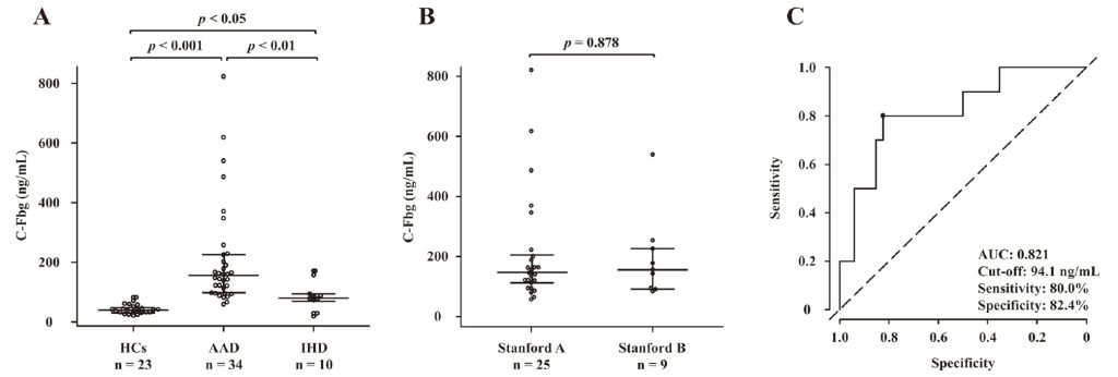
Figure 1 Serum C-Fbg concentrations and ROC analysis results. Initial levels measured of the (A) HCs (n = 23), AAD
patients (n = 34), and IHD patients (n = 10). (B) Levels in patients with Stanford type A and B AAD were compared. The
bar indicates median and interquartile range. The statistical analysis was performed using the Kruskal–Wallis and Steel–Dwass test (A) and the Mann–Whitney U test (B). (C) The ROC analysis of the initial serum C-Fbg level of AAD versus
IHD patients is shown. AAD, acute aortic dissection; C-Fbg, citrullinated fibrinogen; HCs, healthy controls; IHD, ischemic heart disease; ROC, receiver operating characteristic
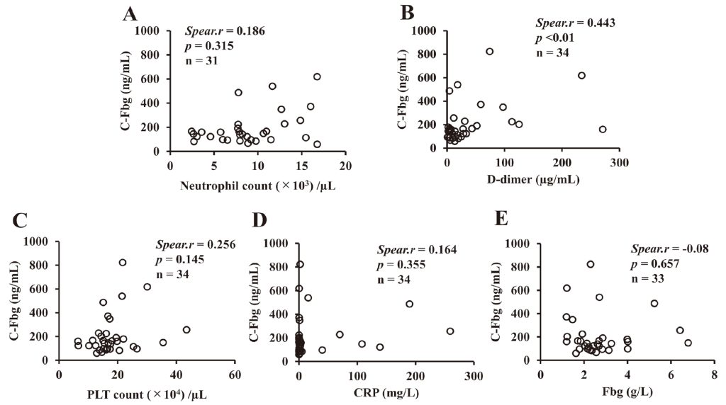
Figure 2 Correlation between C-Fbg concentration and inflammatory markers in the initial sample taken from patients
with AAD. The correlations between C-Fbg and neutrophil count (A), D-dimer (B), PLT count (C), CRP level (D), and
Fbg concentration (E) are shown. The significance of the correlation (p) was analyzed with Spearman’s rank correlation
coefficient (Spear. r). AAD, acute aortic dissection; C-Fbg, citrullinated fibrinogen; CRP, C-reactive protein; Fbg, fibrinogen; PLTc platelet count
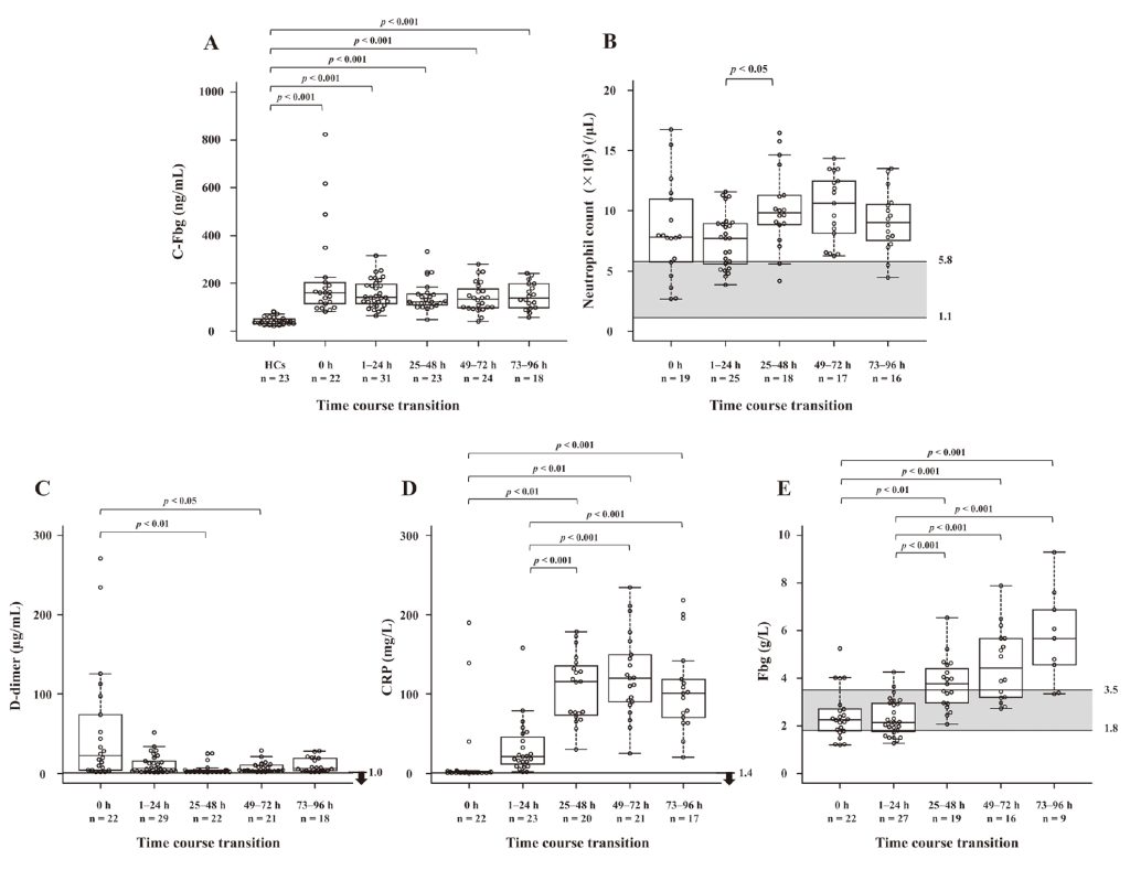
Figure 3 Time course of C-Fbg and other inflammatory markers. Samples from 23 AAD patients were classified by the
submission time; 0 h (the first submission sample), 1-24 h, 25-48 h, 49-72 h, 73-96 h. The concentration of serum C-Fbg
(A), neutrophil counts (B), and concentrations of D-dimer (C), CRP (D), and Fbg (E) are shown. The bar indicates the
medians with the interquartile range. The reference interval of each marker at Shinshu University Hospital is depicted by a gray area or bar and arrow. The statistical analyses were performed by the Kruskal–Wallis and Steel–Dwass test. AAD, acute aortic dissection; C-Fbg, citrullinated fibrinogen; CRP, C-reactive protein; Fbg, fibrinogen; HCs, healthy controls
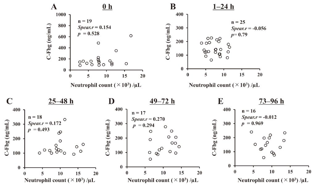
Figure 4 Correlation between C-Fbg concentration and neutrophil count in the sample of patients with AAD at each
classified phase as follows: 0 h (initial sample) (A), 1–24 h (B), 25–48 h (C), 49–72 h (D), and 73–96 h (E) after
admission. The significance of the correlation (p) was analyzed with Spearman’s rank correlation coefficient (Spear. r).
AAD, acute aortic dissection; C-Fbg, citrullinated fibrinogen
Funding
This work was supported by the Charitable Trust Laboratory Medicine Research Foundation of Japan [2019] and JSPS KAKENHI (no. JP20K07822).
Authorship Contributions
YH, AI, TH, NO, and SF designed the study and prepared the manuscript. SF, TI, YH, and FT performed the experiments and analyzed the data. TI, YU, MY, YH, and SF collected the samples and data. All authors have read and approved the final version of the manuscript.
Disclosure of Conflicts of Interest
All authors declare no conflicts of interest.
References
- Luo F, Zhou XL, Li JJ, et al. Inflammatory response is associated with aortic dissection. Ageing Res Rev 2009; 8(1):31-5. doi: 10.1016/j.arr.2008.08.001.PubMed
- Ogino H, Iida O, Akutsu K, et al. JCS/JSCVS/JATS/JSVS Guideline on Diagnosis and Treatment of Aortic Aneurysm and Aortic Dissection. 2020. (in Japanease) Available from: URL: https://www.j-circ.or.jp/cms/wp-content/uploads/2020/07/JCS2020_Ogino.pdf (Accessed December 24, 2022).PubMed
- Cifani N, Proietta M, Tritapepe L, et al. Stanford-A acute aortic dissection, inflammation, and metalloproteinases: A review. Ann Med 2015; 47(6):441-6. doi: 10.3109/07853890.2015.1073346.PubMed
- Niinimäk E, Pynnönen V, Kholova I, et al. Neovascularization with chronic inflammation characterizes ascending aortic dissection. Anatol J Cardiol 2018; 20(5):289-95. doi: 10.14744/AnatolJCardiol.2018.42223.PubMed
- Wu D, Choi JC, Sameri A, et al. Inflammatory cell infiltrates in acute and chronic thoracic aortic dissection. Aorta (Stamford) 2013; 1(6): 259-67. doi: 10.12945/j.aorta.2013.13-044.PubMed
- Son BK, Sawaki D, Tomida S, et al. Granulocyte macrophage colony-stimulating factor is required for aortic dissection/intramural haematoma. Nat Commun 2015; 6: 6994. doi: 10.1038/ncomms7994.PubMed
- Anzai A, Shimoda M, Endo J, et al. Adventitial CXCL1/G-CSF expression in response to acute aortic dissection triggers local neutrophil recruitment and activation leading to aortic rupture. Circ Res 2015; 116(4):612-23. doi: 10.1161/CIRCRESAHA.116.304918.PubMed
- Xu L, Burke A. Acute medial dissection of the ascending aorta: Evolution of reactive histologic changes. Am J Surg Pathol 2013; 37(8):1275-82. doi: 10.1097/PAS.0b013e318294adc3.PubMed
- Wen D, Zhou XL, Li JJ, Hui RT. Biomarkers in aortic dissection. Clin Chim Acta. 2011; 412(9-10):688-95. doi: 10.1016/j.cca.2010.12.039.
- Nienaber CA, Clough RE. Management of acute aortic dissection. Lancet 2015; 385(9970):800-11. doi: 10.1016/s0140-6736(14)61005-9.PubMed
- Katoh H, Suzuki T, Yokomori K, et al. A novel immunoassay of smooth muscle myosin heavy chain in serum. J Immunol Methods 1995; 185(1): 57-63. doi: 10.1016/0022-1759(95)00104-i.PubMed
- Kurihara T, Shimizu-Hirota R, Shimoda M, et al. Neutrophil-derived matrix metalloproteinase 9 triggers acute aortic dissection. Circulation 2012; 126(25): 3070-80. doi: 10.1161/circulationaha.112.097097.PubMed
- Shinohara T, Suzuki K, Okada M, et al. Soluble elastin fragments in serum are elevated in acute aortic dissection. Arterioscler Thromb Vasc Biol 2003; 23(10): 1839-44. doi: 10.1161/01.PubMed
- Suzuki T, Distante A, Zizza A, et al. Preliminary experience with the smooth muscle troponin-like protein, calponin, as a novel biomarker for diagnosing acute aortic dissection. Eur Heart J 2008; 29(11): 1439-45. doi: 10.1093/eurheartj/ehn162.PubMed
- Suzuki T, Trimarchi S, Sawaki D, et al. Circulating transforming growth factor-beta levels in acute aortic dissection. J Am Coll Cardiol 2011; 58(7): 775. doi: 10.1016/j.jacc.2010.01.079.PubMed
- Akutsu K, Sato N, Yamamoto T et al. A rapid bedside D-dimer assay (cardiac D-dimer) for screening of clinically suspected acute aortic dissection. Circ J 2005; 69(4): 397-403. doi: 10.1253/circj.69.397.PubMed
- Weber T, Högler S, Auer J, et al. D-dimer in acute aortic dissection. Chest 2003; 123(5): 1375-8. doi: 10.1378/chest.123.5.1375.
- Sodeck G, Domanovits H, Schillinger M, et al. D-dimer in ruling out acute aortic dissection: A systematic review and prospective cohort study. Eur Heart J 2007; 28(24): 3067-75. doi: 10.1093/eurheartj/ehm484.PubMed
- Zhang B, Wang Y, Guo J, et al. Nomogram to differentiate between aortic dissection and non-ST segment elevation acute coronary syndrome: a retrospective cohort study. Cardiovasc Diagn Ther 2021; 11(2): 457-66. doi: 10.21037/cdt-20-935.PubMed
- 20) Valesini G, Gerardi MC, Iannuccelli C, et al. Citrullination and autoimmunity. Autoimmun Rev 2015; 14(6): 490-7. doi: 10.1016/j.autrev.2015.01.013.PubMed
- György B, Tóth E, Tarcsa E, et al. Buzás EI. Citrullination: a posttranslational modification in health and disease. Int J Biochem Cell Biol 2006; 38(10): 1662–77. doi: 10.1016/j.biocel.2006.03.008.PubMed
- Chavanas S, Méchin MC, Takahara H, et al. Comparative analysis of the mouse and human peptidylarginine deiminase gene clusters reveals highly conserved non-coding segments and a new human gene, PADI6. Gene 2004; 330: 19-27. doi: 10.1016/j.gene.2003.12.038.PubMed
- Mohanan S, Cherrington BD, Horibata S, et al. Potential role of peptidylarginine deiminase enzymes and protein citrullination in cancer pathogenesis. Biochem Res Int 2012; 2012: 895343. doi: 10.1155/2012/895343.PubMed
- Wegner N, Lundberg K, Kinloch A, et al. Autoimmunity to specific citrullinated proteins gives the first clues to the etiology of rheumatoid arthritis. Immunol Rev 2010; 233(1): 34-54. doi: 10.1111/j.0105-2896.2009.00850.x.PubMed
- Fujimura S, Higuchi Y, Usami Y, et al. Changes in serum citrullinated fibrinogen concentration associated with the phase of bacteremia patients. Clin Chim Acta 2021; 512: 127-34. doi: 10.1016/j.cca.2020.10.038.PubMed
- Zhang J, Jiang Y, Gao C, et al. Risk factors for hospital death in patients with acute aortic dissection. Heart Lung Circ 2015; 24(4): 348-53. doi: 10.1016/j.hlc.2014.10.009.PubMed
- Okumura N, Haneishi A, Terasawa F. Citrullinated fibrinogen shows defects in FPA and FPB release and fibrin polymerization catalyzed by thrombin. Clin Chim Acta. 2009; 401(1-2):119–23. doi: 10.1016/j.cca.2008.12.002.PubMed
- Hirota-Kawadobora M, Kani S, Terasawa F, et al. Functional analysis of recombinant Bbeta15C and Bbeta15A fibrinogens demonstrates that Bbeta15G residue plays important roles in FPB release and in lateral aggregation of protofibrils. J Thromb Haemost 2005; 3(5): 983–90. doi: 10.1111/j.1538-7836.2005.01294.x.PubMed
- Terasawa F, Ishii W, Higuchi Y, et al. Measurement of the serum levels of citrullinated fibrinogen and its antibodies in rheumatoid arthritis patients. Int J Anal Bio-Sci 2016; 39(4): 263–70. (in Japanease)
- Suzuki T, Distante A, Zizza A, et al. Diagnosis of acute aortic dissection by D-dimer: the International Registry of Acute Aortic Dissection Substudy on Biomarkers (IRAD-Bio) experience. Circulation 2009; 119(20): 2702-7. doi: 10.1161/CIRCULATIONAHA.108.833004.PubMed
- Ohlmann P, Faure A, Morel O, et al. Diagnostic and prognostic value of circulating D-Dimers in patients with acute aortic dissection. Crit Care Med 2006; 34(5): 1358-64. doi: 10.1097/01.Ccm.0000216686.72457.Ec.PubMed
- Hazui H, Fukumoto H, Negoro N, et al. Simple and useful tests for discriminating between acute aortic dissection of the ascending aorta and acute myocardial infarction in the emergency setting. Circ J 2005; 69(6): 677-82. doi: 10.1253/circj.69.677.PubMed
- Lahoz-Beneytez J, Elemans M, Zhang Y, et al. Human neutrophil kinetics: Modeling of stable isotope labeling data supports short blood neutrophil half-lives. Blood 2016; 127(26): 3431-8. doi: 10.1182/blood-2016-03-700336.PubMed
- Tsai TT, Nienaber CA, Eagle KA. Acute aortic syndromes. Circulation 2005; 112(24): 3802-13. doi: 10.1161/CIRCULATIONAHA.105.534198.
- Mannucci PM, Duga S, Peyvandi F. Recessively inherited coagulation disorders. Blood 2004; 104(5): 1243-52. doi: 10.1182/blood-2004-02-0595.PubMed
- Yang S, Xiao Y, Du Y, et al. Diagnostic and Prognostic Value of Neutrophil Extracellular Trap Levels in Patients With Acute Aortic Dissection. Front Cardiovasc Med 2021; 8: 683445. doi: 10.3389/fcvm.2021.683445.PubMed
- Pan G, Liao M, Dai Y, et al. Inhibition of sphingosine-1-phosphate receptor 2 prevents thoracic aortic dissection and rupture. Front Cardiovasc Med 2021; 8: 748486. doi: 10.3389/fcvm.2021.748486.PubMed
- Brinkmann V, Reichard U, Goosmann C, et al. Neutrophil extracellular traps kill bacteria. Science 2004; 303(5663): 1532–5. doi: 10.1126/science.1092385.PubMed
- Hasler P, Giaglis S, Hahn S. Neutrophil extracellular traps in health and disease. Swiss Med Wkly 2016; 146: w14352. doi: 10.4414/smw.2016.14352.PubMed
- Darrah E, Andrade F. Rheumatoid arthritis and citrullination. Curr Opin Rheumatol 2018; 30(1): 72-8. doi: 10.1097/bor.0000000000000452.PubMed
- Tomida S, Aizawa K, Nishida N, et al. Indomethacin reduces rates of aortic dissection and rupture of the abdominal aorta by inhibiting monocyte/macrophage accumulation in a murine model. Sci Rep 2019; 9(1): 10751. doi: 10.1038/s41598-019-46673-z.PubMed
- Vossenaar ER, Zendman AJ, van Venrooij WJ, et al. PAD, a growing family of citrullinating enzymes: Genes, features and involvement in disease. BioEssays 2003; 25(11): 1106-18. doi: 10.1002/bies.10357.PubMed
- Peng W, Peng Z, Chai X, et al. Potential biomarkers for early diagnosis of acute aortic dissection. Heart Lung 2015; 44(3): 205-8. doi: 10.1016/j.hrtlng.2015.01.006.PubMed
- Karakoyun S, Gürsoy MO, Akgün T, et al. Neutrophil-lymphocyte ratio may predict in-hospital mortality in patients with acute type A aortic dissection. Herz 2015; 40(4): 716-21. doi: 10.1007/s00059-014-4121-2.PubMed
- Oz K, Iyigun T, Karaman Z, et al. Prognostic Value of neutrophil to lymphocyte ratio and risk factors for mortality in patients with Stanford type A aortic Dissection. Heart Surg Forum 2017; 20(3): E119-23. doi: 10.1532/hsf.1736.PubMed
- Onuk T, Güngör B, Karataş B, et al. increased neutrophil to lymphocyte ratio is associated with in-hospital mortality in patients with aortic dissection. Clin Lab 2015; 61(9): 1275-82.Oz K, Iyigun T, Karaman Z, et al. Prognostic Value of neutrophil to lymphocyte ratio and risk factors for mortality in patients with Stanford type A aortic Dissection. Heart Surg Forum 2017; 20(3): E119-23. doi: 10.1532/hsf.1736.PubMedPubMed
- Li S, Yang J, Dong J, et al. Neutrophil to lymphocyte ratio and fibrinogen values in predicting patients with type B aortic dissection. Sci Rep 2021; 11(1): 11366. doi: 10.1038/s41598-021-90811-5.PubMed