Takashi Mitamura*1, Kenji Oku*2,3, Yuichiro Fujieda*3, Hiroshi Asano*1, Chisa Shimada*4, Ayako Nozaki*5, Daisuke Endo*1, Hidemichi Watari*1
Mitamura T, Oku K, Fujieda Y, Asano H, Shimada C, Nozaki A, Endo D, Watari H . Antiphospholipid antibodies in patients with ovarian cancer: A prospective pilot study. Lab Med Int 2025; 4(3): 91-99. doi: 10.51041/lmi.4.3_91
Report
Lab Med Int 2025; 4(3): 91-99
Department of Obstetrics and Gynecology, Hokkaido University Faculty of Medicine, Hokkaido University, North 15, West 7, Kita-Ku, Sapporo, zip code 0608638, Japan.
Tel: +81-11-706-5941, Fax: +81-11-706-7711
E-mail: takami”@”huhp.hokudai.ac.jp
Received April 13, 2025; accepted May 22, 2025
*1 Department of Obstetrics and Gynecology, Hokkaido University Faculty of Medicine, Hokkaido University, Sapporo, Japan.
*2 Division of Rheumatology, Department of Internal Medicine, Tokai University School of Medicine, Kanagawa, Japan.
*3Department of Rheumatology, Endocrinology and Nephrology Faculty of Medicine and Graduate School of Medicine Hokkaido University, Sapporo, Japan.
*4Division of Gynecologic Oncology, National Hospital Organization, Hokkaido Cancer Center, Sapporo, Japan.
*5Department of Obstetrics and Gynecology, Asahikawa-Kosei General Hospital, Asahikawa, Japan.
ABSTRACT
Background; Ovarian cancer is associated with a high incidence of thromboembolism, and it has been suggested that the mechanisms, particularly when apoptosis is induced by chemotherapy, share common underpinnings with antiphospholipid syndrome (APS) -associated thromboembolism. A clinical study was conducted to elucidate the expression of antiphospholipid antibody (aPL) in patients undergoing various treatment steps.
Methods; Fifteen patients with newly diagnosed ovarian cancer, primary peritoneal cancer, or fallopian tube cancer were prospectively and consecutively enrolled to measure lupus anticoagulants, anti-cardiolipin antibodies, and anti-PS/PT antibodies. The observational period for thrombotic events after blood sampling ranged from 5 to 13 months.
Results; Six patients received systemic chemotherapy as neoadjuvant or adjuvant therapy before blood sampling; four patients had thromboembolic diseases, including cerebral infarction; and four patients had clear cell carcinoma. None of the patients showed aPL, regardless of chemotherapy induction. Univariate analysis showed no major clinical characteristics (advanced age, history of thromboembolisms, clear cell carcinoma, large tumor diameter, high body mass index, and advanced FIGO stage) that correlated with thrombosis, while the histological subtype of clear cell carcinoma was associated with elevated plasma D-dimer levels above 8.45 mg/L (P=0.03).
Conclusions; Based on this pilot study with a limited number of patients, ovarian cancer and its treatment may have no direct association with the induction of APS or aPL, regardless of the clinicopathological background or induction of chemotherapy with cytotoxic agents. Further research in this area is warranted.
〔Lab Med Int 2025; 4(3): 91-99〕
Key Words
Ovarian Cancer, Antiphospholipid Syndrome, Antiphospholipid Antibody, Chemotherapy
I. Introduction
Cancer-associated thrombosis is the second leading cause of mortality among cancer patients following disease progression1). The risk of thromboembolism in individuals with cancer is intricately linked to tumor characteristics, including primary site, histological grade, or tumor node metastasis stage, as well as to cancer treatments, such as surgery, hospitalization, central venous catheter placement, systemic chemotherapy, radiotherapy, anti-angiogenesis agents, immunomodulatory drugs, hormonal therapy, erythropoiesis-stimulating agents, and red blood cell or platelet transfusions1). The mechanisms underlying cancer-associated thrombosis are multifaceted, involving direct activation of platelets by cancer cells and procoagulant molecules such as tissue factor (TF) and P-selectin derived from tumor cell microvesicles2). These pathways illustrate the complex interplay between malignancy and hypercoagulability.
Antiphospholipid syndrome (APS) is a systemic autoimmune condition characterized by arterial, venous, or microvascular thrombosis, recurrent pregnancy loss, and non-thrombotic manifestations in the presence of persistent anti-phospholipid antibodies (aPLs)3). The pathogenic role of aPLs is well documented. These autoantibodies induce a prothrombotic state primarily through the activation of vascular endothelial cells and monocytes, resulting in increased TF production4). This mechanism, known as the procoagulant cell activation theory, underscores the pivotal role of blood-borne TF in APS-associated hypercoagulability. The representative aPL anti-cardiolipin antibodies are auto-antibodies that target plasma proteins, primarily β2-glycoprotein I (GPI), which binds to anionic phospholipids5)6). Phosphatidylserine-dependent anti-prothrombin antibodies (aPS/PTs) exhibit structural characteristics similar to those of antiphospholipid antibodies by binding to prothrombin, which undergoes conformational changes upon interaction with negatively charged phospholipids. These antibodies also possess prothrombotic properties, and are increasingly recognized as novel or alternative antiphospholipid antibodies, potentially substituting lupus anticoagulants (LACs) in certain diagnostic contexts4)7). Emerging evidence suggests a potential association between APS and thrombosis in cancer patients, possibly mediated by the overexpression of phosphatidylserine on cell surfaces, including in ovarian cancer8)-11). Although the precise mechanisms remain elusive and may vary by cancer type, this link warrants further exploration.
Ovarian cancer is the most lethal malignancy of the female reproductive system, claiming the lives of over 200,000 women each year12). The standard treatment is debulking surgery to achieve no gross residual tumor, followed by adjuvant platinum-based chemotherapy in both primary and recurrent disease13). In addition, several clinical trials demonstrated that the anti-angiogenesis agent bevacizumab improved the prognosis of patients with advanced14) and recurrent ovarian cancer15), and combination drug therapy with bevacizumab is currently used as a control arm to develop novel treatments in randomized clinical trials16). Notably, ovarian cancer has a disproportionately high incidence of thromboembolism relative to other malignancies17)18) and has been associated with Trousseau syndrome as a cerebral infarction19). Chemotherapy has been implicated in the externalization of negatively charged phospholipids, such as phosphatidylserine20), due to apoptosis in ovarian cancer21), further increasing the thrombotic risk. The shared pathophysiological underpinnings of APS-associated thrombosis and cancer-associated thrombosis, particularly the formation of fibrin clots, suggest a potential overlap between these conditions22). This prompted our hypothesis that aPLs may be more frequently detected in patients with ovarian cancer. Recognizing the critical importance of anticipating thrombotic events and instituting appropriate prophylactic measures, we explored the utility of aPL testing. However, the clinical relevance of aPLs in ovarian cancer remains controversial, as the current data are limited to case reports and small-scale studies. To address this gap, we conducted a prospective study to elucidate the correlation between the expression of aPL and clinical thrombotic events, with a focus on their timing relative to the administration of chemotherapy. Through this investigation, we sought to advance the understanding of thrombotic risk in ovarian cancer and refine strategies for its management.
II. Materials and methods
Ovarian Cancer Patients
From 2016 to 2017, we consecutively invited eligible Japanese patients from Hokkaido University Hospital and its affiliated hospitals to participate in our clinical study. These patients were histologically diagnosed with borderline epithelial ovarian, ovarian, primary peritoneal, or fallopian tube cancer, as confirmed by certified pathologists, irrespective of their clinical characteristics. Body mass index (BMI), history of thromboembolisms before the initial diagnosis of each patient’s gynecological tumors, chemotherapy before blood sampling, disease stage, tumor diameter, and disease status were confirmed at registration. All thromboembolic events associated with the diagnosis of each patient’s gynecological tumors were confirmed 5 months after the last subjects were registered in November 2017. Thromboembolisms were assessed when they were suspected by ultrasonography or contrast-enhanced computed tomography at the physicians’ choice, and we did not perform routine screenings. Certified radiologists checked all the results of imaging tests to diagnose thromboembolism. D-dimer levels were checked in all patients at the same time as blood sampling for this study.
This study was conducted according to the 1964 Declaration of Helsinki. The Institutional Review Board of Hokkaido University approved all experiments on the human genome for this study (Registration ID: 014-0106). Plasma and serum samples were prospectively collected from the patients, and antiphospholipid antibodies were measured as follows. Samples were collected in tubes containing a one-tenth volume of 0.105M sodium citrate and centrifuged immediately at 4℃. Plasma samples were depleted of platelets by filtration and stored at −80℃.
Antiphospholipid antibody (aPL) tests
Clotting tests to determine the activated partial thromboplastin time (aPTT) and dilute Russell’s viper venom time (dRVVT) were performed for LAC determination using a semiautomated hemostasis analyzer (STart 4; Diagnostica Stago) according to the guidelines recommended by the Subcommittee on Lupus Anticoagulant/Antiphospholipid Antibody of the Scientific and Standardization Committee of the International Society on Thrombosis and Hemostasis 23. IgG and IgM anti-cardiolipin antibodies (aCL) were assayed using a standard enzyme-linked immunosorbent assay (ELISA)24). Anti-PS/PT antibodies were detected by an ELISA as previously described25).
Statistical analysis
Using a univariate analysis, we analyzed any correlations between the clinical parameters and thrombosis or high D-dimer values and set P values of 0.05 as being statistically significant for Fisher’s exact test. All statistical analyses were performed using the JMP® Pro software program (ver. 14.0.0; SAS Institute, Cary, NC, USA).
III. Results
Disease characteristics of the study patients
We prospectively enrolled 15 patients with newly diagnosed ovarian, primary peritoneal, or fallopian tube cancer. All approached patients agreed to participate, resulting in complete consecutive enrollment. The clinical characteristics of the patients are summarized in Table 1. The median age and BMI at registration were 63 years (range 41 – 82) and 23.4 kg/m2 (range 16.1 – 34.3), respectively. Blood samples were collected either before or during treatment for the primary disease, except for one patient with recurrent disease. None of the patients had a history of thromboembolism unrelated to their current treatments for gynecological tumors, nor did they have untreated severe hypertension, diabetes, or hyperlipidemia. Two patients in the cohort were smokers; neither had experienced thromboembolism before blood sampling was conducted. None of our patients used pro-coagulant drugs at the time of blood sampling. We histologically diagnosed gynecological tumors using surgical specimens in 14 patients (93.3%) and a pleural effusion cell block in one patient (6.7%). The distribution of histological subtypes among the patients was as follows: serous carcinoma (33.3%), clear cell carcinoma (26.7%), mucinous carcinoma (20.0%), and endometrioid carcinoma (13.3%). Forty percent of patients (n=6) received systemic chemotherapy as neoadjuvant or adjuvant therapy before blood sampling. The median number of chemotherapy cycles was 3 (range, 1–7), and the regimen included tri-weekly paclitaxel and carboplatin (TC), dose-dense TC, tri-weekly TC with bevacizumab, gemcitabine as a single agent, and a combination of irinotecan and nedaplatin. Figure 1 illustrates each patient’s treatment course and the timing of the blood sampling. One patient (no.2 in Figure 1) received chemotherapy with bevacizumab before blood sampling. None of the patients had previously undergone radiotherapy. The follow-up period for thrombotic events after study registration ranged from 5 to 13 months. Three patients had preoperative deep venous thrombosis (DVT). In these three patients, blood samples were obtained from two patients before the diagnosis of DVT. In the other patient, we obtained a blood sample after the diagnosis of DVT, and this patient was not taking anti-coagulant drugs because the thrombi were organized. In addition, another patient with systemic thrombosis and cerebral infarction was diagnosed with Trousseau’s syndrome and was taking warfarin during blood sampling for this study. The remaining 11 patients either had no thromboembolisms (n=8) or had temporal operation-related DVT (n=3).
Results of blood examinations and aPLs
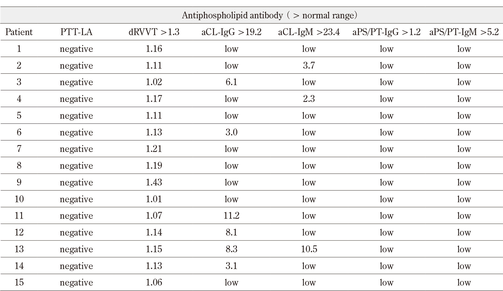
None of the patients tested positive for syphilis at the time of the diagnosis of the gynecological tumor. The D-dimer level at study registration ranged from 0.5 to 24.96 mg/L, and 10 of 15 patients (66.7%) were positive when we set the cutoff value to the general threshold up to 1.0 mg/L. Regarding aPLs, only one patient was positive for the dRVVT screening test (Table 2). However, this patient was negative for all the other screening tests (no.9 in Figure 1). Therefore, none of the patients–including one with cerebral infarction of Trousseau’s syndrome and six who were registered after chemotherapy–showed the presence of aPLs (Table 2). We subsequently asked 5 of the 15 (33.3%) patients to provide additional blood samples from 1 to 6 months after the first sampling, regardless of the timing of cancer treatment, and no patients had aPLs again (not shown in Figure 1). We also measured the expression of aPLs in four patients with mucinous borderline ovarian tumors using the same methods, and none of these patients had aPLs (data not shown).
A univariate analysis for risk factors of elevated D-dimer
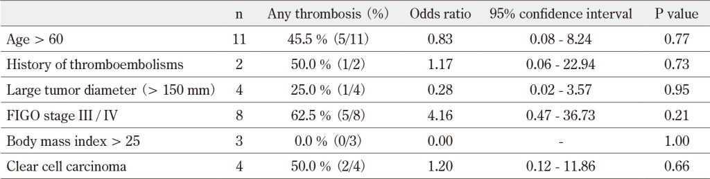
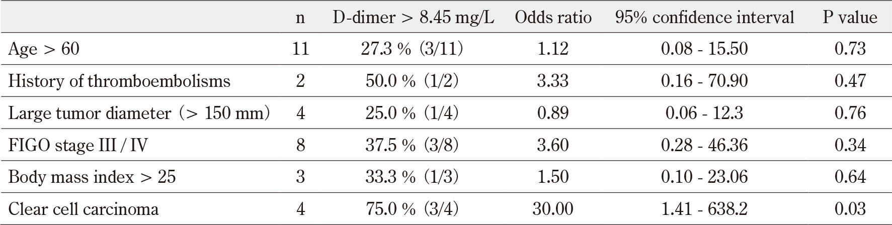
Age (> 60 years), history of thromboembolisms, large tumor diameter (> 150 mm), high body mass index (> 25), histological subtypes of clear cell carcinoma, and advanced FIGO stage (III of IV) were not significantly correlated with thrombosis during the study period. These factors were also not significantly correlated with an elevated plasma D-dimer level at study registration when we set the cutoff value to the general threshold up to 1.0 mg/L. On the other hand, when we set the cutoff value of D-dimer to 8.45 mg/L, which was the mean value of acute phase patients with Trousseau’s syndrome in a previous study 26, the histological subtype of clear cell carcinoma was significantly correlated with an elevated plasma D-dimer level (P = 0.03, Table 3). Although the limited sample size and heterogeneity of clinical backgrounds preclude definitive conclusions regarding the correlation between thrombotic events and prognosis, the 5-year overall survival rate was comparable at 57.1% among patients with and without thrombotic events, except for patient 4 in Figure 1, whose prognosis remains unknown.
Table 1 Disease characteristics and antiphospholipid antibody expression of the study patients (n=15)
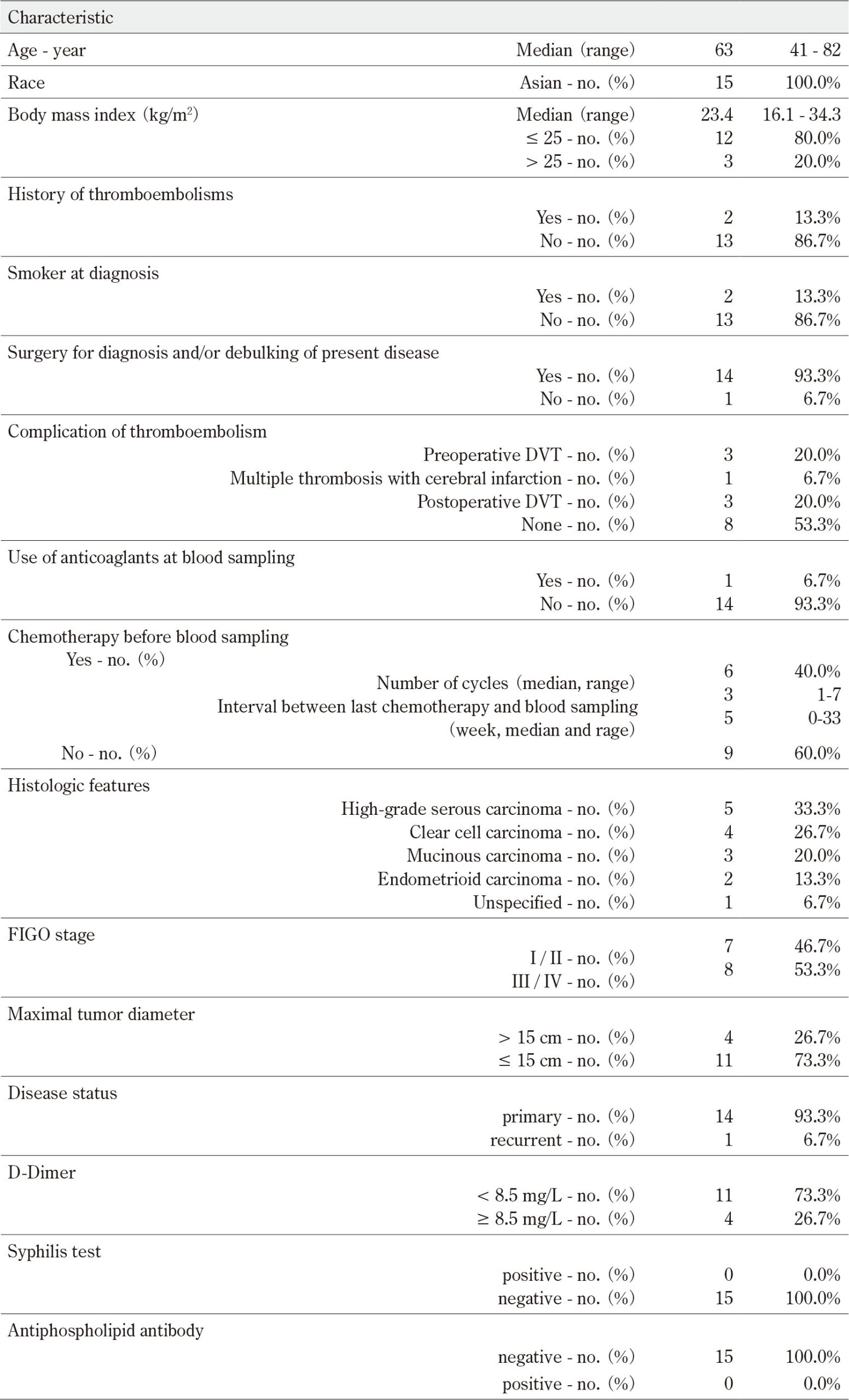
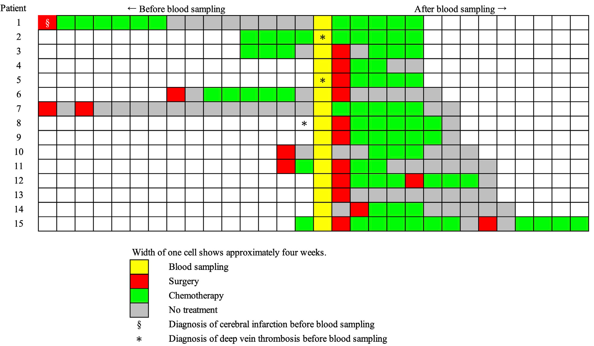
Figure 1 Clinical course and the timing of blood sampling
Table 2 Results of antiphospholipid antibodies (n=15)
IV. Discussion
To our knowledge, this study included the largest cohort of ovarian cancer patients to be investigated in this context. Clinically significant thrombotic events, excluding postoperative DVT, a common complication of abdominal surgery, were observed in 4 of the 15 patients. This finding aligns with existing reports indicating a high incidence of thrombotic events in ovarian cancer. The lack of detectable aPL in ovarian cancer patients in this study may be explained by several factors. First, ovarian cancer cells, whether treatment-naïve or following chemotherapy, might not prominently expose negatively charged phospholipids on their cell surfaces, which are essential for aPL production. Second, even if such phospholipids are expressed on cell surfaces, additional genetic predispositions associated with autoimmune diseases may be necessary to trigger the generation of aPL. Further basic and clinical investigations are required to confirm these hypotheses. Finally, the current finding–that ovarian cancer, despite its strong association with thrombosis, exhibits minimal aPL expression–raises the possibility that aPLs may have relatively limited involvement in cancer-associated thrombosis in other malignancies as well. However, this remains to be validated. This cohort included four patients with clear cell carcinoma, a rare histologic subtype in Western countries. This subtype is reported to carry the highest risk of serious thromboembolism among ovarian carcinomas19), probably due to an excessive production of tissue factor27). Although our study was unable to demonstrate the risk factors for thromboembolism in patients with ovarian cancer due to the small number of subjects, the findings of our univariate analysis did suggest a correlation between clear cell carcinoma and elevated plasma D-dimer levels which strongly correlate with an increased risk of thrombosis, particularly venous thromboembolism, in ovarian cancer patients28).
Previous studies have shown that treatment with chemotherapy is an independent risk factor for symptomatic and incidental venous thromboembolism in patients with endometrial, cervical, ovarian, tubal, or peritoneal cancer29)30). In addition, it has been suggested that chemotherapy, which is a mainstay in the treatment of ovarian cancer, may induce aPL via the externalization of negatively charged phospholipids due to apoptosis. Regarding elevated anionic phospholipid expression associated with tissue damage, aPLs are known to transiently appear during acute infections such as COVID-1931). Therefore, we hypothesized that aPLs could potentially be expressed at various stages in ovarian cancer, where prolonged tumor growth or treatment-related modifications may induce high anionic phospholipid expression levels. However, our study found no significant correlation between aPLs and thromboembolic events in general patients with ovarian cancer, and no aPL induction by chemotherapy with cytotoxic agents. Therefore, with the exception of infrequent situations suspicious for serious catastrophic anti-phospholipid syndrome–characterized by the rapid chronological development of fulminant thrombotic complications leading to multi-organ failure32) –clinicians do not need to routinely check aPLs during chemotherapy with cytotoxic agents in patients with ovarian cancer. However, we cannot discuss the influence of bevacizumab because of the small number of patients.
Although aPLs are generally reported to be present in 1–12% of healthy individuals 31, no positive cases were identified among the 15 patients in this cohort. The presence of aPLs has been proposed to be a potential risk factor for malignancy and mortality. A previous study demonstrated a 2.6-fold increase in the risk of cancer-related mortality in patients positive for anti-cardiolipin antibodies33). Additionally, a prospective study of 1,000 APS patients with APS identified malignancies as one of the most frequent causes of death in this population34). A recent large-scale study on obstetric APS (n=517) reported a significant association between aPL positivity and an increased incidence of malignancy after long-term follow-up (hazard ratio, 2.22), although no ovarian cancer cases were observed35). However, the frequency of aPL induction in ovarian cancer remains poorly understood. Few case reports have documented patients with ovarian cancer who incidentally test positive for aPLs36)- 38). Fewer reports suggest that APS, characterized by the rapid onset of severe thromboembolisms, may arise as a paraneoplastic syndrome in ovarian cancer36)39). For other malignancies, prior research reported an aPL positivity rate of 5.7% among patients with various solid tumors, with higher rates observed in VTE-positive colon cancer (11.3%), breast cancer (7.9%), head and neck cancer (7.7%), and lung cancer (4.7%)40). However, it found no significant difference in thrombosis-free survival between aPL-positive and aPL-negative patients41). Data specific to ovarian cancer were notably absent from these reports, suggesting that aPL positivity may remain infrequent in patients with ovarian cancer.
The tumor microenvironment of ovarian cancer is suggested to be procoagulant, where tumor cell-induced activated platelets interact with endothelial cells, pericytes, mesenchymal stem cells, cancer-associated fibroblasts, adipocytes, immune cells, and extracellular matrix elements through direct interactions or by releasing various modulatory factors and platelet microparticles42). In this study, we did not evaluate intratumoral thrombosis induced by procoagulant factors other than aPLs and its association with systemic thrombosis, including pre-operative DVT or cerebral infarction, and further study is needed. Our study showed that aPLs are not generally associated with systemic venous and arterial thrombosis.
Antiphospholipid antibody detection encompasses various types (including lupus anticoagulant), which require multiple tests for confirmation. Another key marker, aPS/PT, is a frequently observed and characteristic autoantibody of APS; however, its diagnostic utility has only recently been established. This study was associated with the common limitations of antiphospholipid antibody testing. However, based on this pilot study with a limited number of patients and previous studies, ovarian cancer and its treatment may have no direct association with the induction of APS or aPL. Further research in this area is warranted.
Fundings
YF has received research grants from MEDICAL & BIOLOGICAL LABORATORIES CO., LTD. TM is supported by Japan Society for the Promotion of Science (JSPS KAKENHI Grant Number JP 25K10483).
Acknowledgements
We thank Olga Amengual and Tatsuya Atsumi (Department of Rheumatology, Endocrinology and Nephrology Faculty of Medicine and Graduate School of Medicine Hokkaido University, Sapporo, Japan) for supporting this study.
Authorship Contributions
Takashi Mitamura (Conceptualization; Data curation; Formal analysis; Investigation; Methodology; Project administration; Validation; Writing – original draft), Kenji Oku (Conceptualization; Data curation; Formal analysis; Investigation; Methodology; Project administration; Supervision; Validation; Writing – original draft), Yuichiro Fujieda (Methodology; Supervision; Validation; Writing review & editing), Hiroshi Asano (Data curation; Formal analysis; Investigation; Supervision; Validation; Writing – review & editing), Chisa Shimada (Data curation; Investigation; Writing – review & editing), Ayako Nozaki (Investigation; Validation; Writing – review & editing), Daisuke Endo (Investigation; Writing – review & editing), Hidemichi Watari (Supervision; Writing – review & editing).
Disclosure of Conflicts of Interest
No potential conflict of interest relevant to this article was reported.
Table 3 Univariate analysis of the factors associated with high D-dimer values
Univariate analysis of the factors associated with thrombosis
Univariate analysis of the factors associated with high D-dimer values
References
- Farge D, Frere C, Connors JM, et al. 2019 international clinical practice guidelines for the treatment and prophylaxis of venous thromboembolism in patients with cancer. Lancet Oncol. 2019; 20(10): e566-81. doi: 10.1016/S1470-2045(19)30336-5PubMed
- Palacios-Acedo AL, Langiu M, Crescence L, et al. Platelet and Cancer-Cell Interactions Modulate Cancer-Associated Thrombosis Risk in Different Cancer Types. Cancers (Basel). 2022; 14(3): 730. doi: 10.3390/cancers14030730PubMed
- Barbhaiya M, Zuily S, Naden R, et al. The 2023 ACR/EULAR Antiphospholipid Syndrome Classification Criteria. Arthritis Rheumatol. 2023; 75(10): 1687-702. doi: 10.1002/art.42624PubMed
- Oku K, Amengual O, Zigon P, et al. Essential role of the p38 mitogen-activated protein kinase pathway in tissue factor gene expression mediated by the phosphatidylserine-dependent antiprothrombin antibody. Rheumatology (Oxford). 2013; 52(10): 1775-84. doi: 10.1093/rheumatology/ket234PubMed
- Preetam S, Pandey A, Mishra R, et al. Phosphatidylserine: Paving the way for a new era in cancer therapies. Materials Advances. 2024; 5(21): 8384-403. doi: 10.1039/D4MA00511B
- Brusch A. The Significance of Anti-Beta-2-Glycoprotein I Antibodies in Antiphospholipid Syndrome. Antibodies (Basel). 2016; 5(2): 16. doi: 10.3390/antib5020016PubMed
- Wei J, Fujieda Y, Fujita Y, et al. Phosphatidylserine-Dependent Anti-prothrombin Antibodies as a Key Predictor for Systemic Lupus Erythematosus in Patients with Primary Antiphospholipid Syndrome: A retrospective longitudinal cohort study. Mod Rheumatol. 2024; 5(2): 300-6. doi: 10.1093/mr/roae073PubMed
- Zhang J, Dai Z, Yan C, et al. Blocking antibody-mediated phosphatidylserine enhances cancer immunotherapy. J Cancer Res Clin Oncol. 2021; 147(12): 3639-51. doi: 10.1007/s00432-021-03792-3PubMed
- Belzile O, Huang X, Gong J, et al. Antibody targeting of phosphatidylserine for the detection and immunotherapy of cancer. Immunotargets Ther. 2018; 7: 1-14. doi: 10.2147/ITT.S134834PubMed
- Kelleher RJ Jr, Balu-Iyer S, Loyall J, et al. Extracellular Vesicles Present in Human Ovarian Tumor Microenvironments Induce a Phosphatidylserine-Dependent Arrest in the T-cell Signaling Cascade. Cancer Immunol Res. 2015; 3(11): 1269-78. doi: 10.1158/2326-6066.CIR-15-0086PubMed
- Dong HP, Holth A, Kleinberg L, et al. Evaluation of cell surface expression of phosphatidylserine in ovarian carcinoma effusions using the annexin-V/7-AAD assay: clinical relevance and comparison with other apoptosis parameters. Am J Clin Pathol. 2009; 132(5): 756-62. doi: 10.1309/AJCPAVFA8J3KHPRSPubMed
- Cabasag CJ, Fagan PJ, Ferlay J, et al. Ovarian cancer today and tomorrow: A global assessment by world region and Human Development Index using GLOBOCAN 2020. Int J Cancer. 2022; 151(9): 1535-41. doi: 10.1002/ijc.34002PubMed
- Komiyama S, Katabuchi H, Mikami M, et al. Japan Society of Gynecologic Oncology guidelines 2015 for the treatment of ovarian cancer including primary peritoneal cancer and fallopian tube cancer. Int J Clin Oncol. 2016; 21(3): 435-46. doi: 10.1007/s10147-016-0985-xPubMed
- Burger RA, Brady MF, Bookman MA, et al. Incorporation of bevacizumab in the primary treatment of ovarian cancer. N Engl J Med. 2011; 365(26): 2473-83. doi: 10.1056/NEJMoa1104390PubMed
- Pujade-Lauraine E, Hilpert F, Weber B, et al. Bevacizumab combined with chemotherapy for platinum-resistant recurrent ovarian cancer: The AURELIA open-label randomized phase III trial. J Clin Oncol. 2014; 32(13): 1302-8. doi: 10.1200/JCO.2013.51.4489PubMed
- Ray-Coquard I, Pautier P, Pignata S, et al. Olaparib plus Bevacizumab as First-Line Maintenance in Ovarian Cancer. N Engl J Med. 2019; 381(25): 2416-28. doi: 10.1056/NEJMoa1911361PubMed
- Sun MY, Bhaskar SMM. Venous Thromboembolism in Cancer Patients Undergoing Chemotherapy: A Systematic Review and Meta-Analysis. Diagnostics (Basel). 2022; 12(12): 2954. doi: 10.3390/diagnostics12122954PubMed
- Ye L, Cai L, Fu Y, et al. The prevalence, risk factors, and prognostic value of venous thromboembolism in ovarian cancer patients receiving chemotherapy: a systematic review and meta-analysis. World J Surg Oncol. 2021; 19(1): 12. doi: 10.1186/s12957-020-02101-5PubMed
- Takano H, Nakajima K, Nagayoshi Y, et al. Clinical associations of Trousseau’s syndrome associated with cerebral infarction and ovarian cancer. J Gynecol Oncol. 2018; 29(5): e67. doi: 10.3802/jgo.2018.29.e67PubMed
- Segawa K, Nagata S. An Apoptotic ‘Eat Me’ Signal: Phosphatidylserine Exposure. Trends Cell Biol. 2015; 25(11): 639-50. doi: 10.1016/j.tcb.2015.08.003PubMed
- Binju M, Amaya-Padilla MA, Wan G, et al. Therapeutic Inducers of Apoptosis in Ovarian Cancer. Cancers (Basel). 2019; 11(11): 1786. doi: 10.3390/cancers11111786PubMed
- Zabczyk M, Undas A. Fibrin Clot Properties in Cancer: Impact on Cancer-Associated Thrombosis. Semin Thromb Hemost. 2024; 50(3): 402-12. doi: 10.1055/s-0043-1770364PubMed
- Devreese KMJ, de Groot PG, de Laat B, et al. Guidance from the Scientific and Standardization Committee for lupus anticoagulant/antiphospholipid antibodies of the International Society on Thrombosis and Haemostasis: Update of the guidelines for lupus anticoagulant detection and interpretation. J Thromb Haemost. 2020; 18(11): 2828-39. doi: 10.1111/jth.15047PubMed
- Harris EN, Gharavi AE, Patel SP, et al. Evaluation of the anti-cardiolipin antibody test: report of an international workshop held 4 April 1986. Clin Exp Immunol. 1987; 68(1): 215-22. PubMed
- Atsumi T, Ieko M, Bertolaccini ML, et al. Association of autoantibodies against the phosphatidylserine-prothrombin complex with manifestations of the antiphospholipid syndrome and with the presence of lupus anticoagulant. Arthritis Rheum. 2000; 43(9): 1982-93. doi: 10.1002/1529-0131(200009)43:9<1982::AID-ANR9>3.0.CO;2-2PubMed
- Ito S, Kikuchi K, Ueda A, et al. Changes in Serial D-Dimer Levels Predict the Prognoses of Trousseau’s Syndrome Patients. Front Neurol. 2018; 9: 528. doi: 10.3389/fneur.2018.00528PubMed
- Uno K, Homma S, Satoh T, et al. Tissue factor expression as a possible determinant of thromboembolism in ovarian cancer. Br J Cancer. 2007; 96(2): 290-5. doi: 10.1038/sj.bjc.6603552PubMed
- Ye S, Zhang W, Yang J, et al. Pattern of Venous Thromboembolism Occurrence in Gynecologic Malignancy: Incidence, Timing, and Distribution a 10-Year Retrospective Single-institutional Study. Medicine (Baltimore). 2015; 94(50): e2316. doi: 10.1097/MD.0000000000002316PubMed
- Takahashi Y, Fujiwara H, Yamamoto K, et al. Incidence and risk factors for venous thromboembolism in gynecological cancer: the GOTIC-VTE trial. J Thromb Thrombolysis. 2025; 58(2): 299-308. doi: 10.1007/s11239-024-03055-1PubMed
- Heit JA. Predicting the risk of venous thromboembolism recurrence. Am J Hematol. 2012; 87 Suppl 1(Suppl 1): S63-7. doi: 10.1002/ajh.23128PubMed
- Tung ML, Tan B, Cherian R, et al. Anti-phospholipid syndrome and COVID-19 thrombosis: connecting the dots. Rheumatol Adv Pract. 2021; 5(1): rkaa081. doi: 10.1093/rap/rkaa081PubMed
- Miesbach W. Malignancies and catastrophic anti-phospholipid syndrome. Clin Rev Allergy Immunol. 2009; 36(2-3): 91-7. doi: 10.1007/s12016-008-8101-2PubMed
- Endler G, Marsik C, Jilma B, et al. Anti-cardiolipin antibodies and overall survival in a large cohort: preliminary report. Clin Chem. 2006; 52(6): 1040-4. doi: 10.1373/clinchem.2005.063925PubMed
- Cervera R, Khamashta MA, Shoenfeld Y, et al. Morbidity and mortality in the antiphospholipid syndrome during a 5-year period: a multicentre prospective study of 1000 patients. Ann Rheum Dis. 2009; 68(9): 1428-32. doi: 10.1136/ard.2008.093179PubMed
- Gris JC, Mousty E, Bouvier S, et al. Increased incidence of cancer in the follow-up of obstetric antiphospholipid syndrome within the NOH-APS cohort. Haematologica. 2020; 105(2): 490-7. doi: 10.3324/haematol.2018.213991PubMed
- Ideguchi H, Ohno S, Ueda A, et al. Catastrophic antiphospholipid syndrome associated with malignancies (case report and review of the literature). Lupus. 2007; 16(1): 59-64. doi: 10.1177/0961203306073166PubMed
- Ozaki M, Ogata M, Yokoyama T, et al. Prevention of thrombosis with prostaglandin E1 in a patient with catastrophic antiphospholipid syndrome. Can J Anaesth. 2005; 52(2): 143-7. doi: 10.1007/BF03027719PubMed
- Islam MA. Antiphospholipid antibodies and antiphospholipid syndrome in cancer: Uninvited guests in troubled times. Semin Cancer Biol. 2020; 64: 108-13. doi: 10.1016/j.semcan
cer.2019.07.019PubMed - Ruffatti A, Aversa S, Del Ross T, et al. Antiphospholipid antibody syndrome associated with ovarian cancer. A new paraneoplastic syndrome? J Rheumatol. 1994; 21(11): 2162-3. PubMed
- Font C, Vidal L, Espinosa G, et al. Solid cancer, antiphospholipid antibodies, and venous thromboembolism. Autoimmun Rev. 2011; 10(4): 222-7. doi: 10.1016/j.autrev.2010.10.006PubMed
- Bazzan M, Montaruli B, Vaccarino A, et al. Presence of low titre of antiphospholipid antibodies in cancer patients: a prospective study. Intern Emerg Med. 2009; 4(6): 491-5. doi: 10.1007/s11739-009-0316-6PubMed
- Oncul S, Cho MS. Interactions between Platelets and Tumor Microenvironment Components in Ovarian Cancer and Their Implications for Treatment and Clinical Outcomes. Cancers (Basel). 2023; 15(4): 1282. doi: 10.3390/cancers15041282PubMed