Ayumu Kurihara*1,2, Tadakazu Ao*3, Hiroaki Takeo*1, Susumu Matsukuma*2
Kurihara A, Ao T, Takeo H, Matsukuma S. Acute amebic appendicitis associated with relapsing amebic infection: a case report. Lab Med Int 2025; 4(1): 3-7: 125-134. doi: 10.51041/lmi.4.1_3
Case Report
Lab Med Int 2025; 4(1): 3-7
†Corresponding author: Ayumu Kurihara, E-mail: doc41029″@”ndmc.ac.jp
Received May 16, 2024; accepted September 6, 2024
*1 Department of Pathology, Japan Self-Defense Forces Central Hospital, Tokyo, Japan.
*2 Department of Pathology and Laboratory Medicine, National Defense Medical College, Saitama, Japan.
*3 Department of Surgery, Japan Self-Defense Forces Central Hospital, Tokyo, Japan.
ABSTRACT
Acute amebic appendicitis, a rare presentation of Entamoeba histolytica infection, is challenging to distinguish from non-amebic appendicitis because of similarities in symptoms and laboratory data. Here, we describe the case of a 42-year-old Japanese male with amebic appendicitis diagnosed by histopathological examination of the removed appendiceal specimens. In this case, blood eosinophilia was not observed, and amebic trophozoites were histologically distributed not only in surface exudates but also in appendiceal submucosa and muscularis propria. In addition, a thorough interview and additional colonoscopy after the diagnosis revealed a history of amebic colitis and persistent amebic colitis. We believe that physicians should be aware of the possible presence of amebic appendicitis because of its higher mortality rate compared to non-amebic appendicitis.
〔Lab Med Int 2025; 4(1): 3-7〕
Key Words
amebiasis, acute appendicitis, Entamoeba histolytica, amebic colitis, eosinophilia
I. Introduction
The main manifestations of Entamoeba histolytica (E. histolytica) infection are amebic colitis and amebic liver abscess.1) Almost all (93%) cases of amebic colitis involve the ileocecal region; 2) however, amebic appendicitis is rare,2) accounting for only 0.5–2.3% of acute appendicitis cases even in endemic developing countries.3) Amebic appendicitis is frequently overlooked clinically, and its definite diagnosis requires pathological examination of the removed vermiform appendix, thus delaying therapy.4),5) Here, we describe the unique clinicopathological findings of a pertinent case to expand our knowledge of amebic appendicitis.
II. Case report
A 42-year-old Japanese male was brought to our hospital by ambulance because of exacerbating and moving abdominal pain. The patient was an office worker, and had a history of depression. Physical examination revealed localized tenderness in the right lower abdomen, with muscle guarding. Laboratory tests of peripheral blood revealed a white blood cell (WBC) count of 12,080 /μL without eosinophilia (240 /μL) and serum C-reactive protein (CRP) level of 0.49 mg/dL. Serum albumin is 4.6 g/dL and other serological data ruled out human immunodeficiency virus, hepatitis B virus, and hepatitis C virus infection, so there was no evidence of malnutrition or immunodeficiency. Computed tomography revealed a thickened appendiceal wall with contrast enhancement, multiple appendicoliths, and periappendiceal fat stranding. These findings suggested acute appendicitis. Laparoscopy revealed minimal ascites and no peritoneal abscess, and appendectomy was performed. On post-operative day (POD) 3, serum CRP level was slightly elevated but WBC was decreased (2.46 mg/dL and 3,790 /μL, respectively), and on POD 4, the patient was discharged without any complication. On POD5, the appendectomized specimen was pathologically diagnosed as an amebic infection. On POD18, an in-depth medical interview was performed and revealed the patient’s hidden medical history: 1) one year before this episode, the patient had been diagnosed with amebic colitis of entire colon by colonoscopy at another clinic; 2) the patient was treated with 5-days oral metronidazole, his symptoms disappeared, and his follow-up at the clinic was completed. By POD18, serum CRP levels and WBC count were within normal ranges and his symptom was disappeared, so additional treatment seemed unnecessary, but we recommended colonoscopy just in case. On POD49, the patient underwent additional colonoscopy at the clinic, and biopsy specimens from the ileocecal erosive lesions showed a relapsing amebic infection without colitis of any other part. The patient was given a 10-day metronidazole treatment. Six months after treatment, a follow-up colonoscopy revealed complete endoscopic remission, and the patient is alive and well.
III. Histopathological Findings
The removed vermiform appendix showed dark mucosal changes at the tip (Figure 1). Microscopically, a neutrophil-predominant inflammatory infiltrate with a small number of eosinophils, lymphocytes, and plasma cells involved the entire thickness of the appendiceal wall, chiefly located on the appendiceal tip, accompanied by focal ulcers. Hematoxylin and eosin (HE) -stained sections identified oval amebic trophozoites with foamy cytoplasm and eosinophilic nucleus-like structures (Figure 2). These amebic trophozoites are highlighted in bright red by periodic acid-Schiff (PAS) staining (Figure 2b, inset). Amebic trophozoites were predominantly distributed within the appendiceal wall rather than on the surface (Figure 3). Trophozoites phagocytosed erythrocytes in the cytoplasm suggest active inflammation.
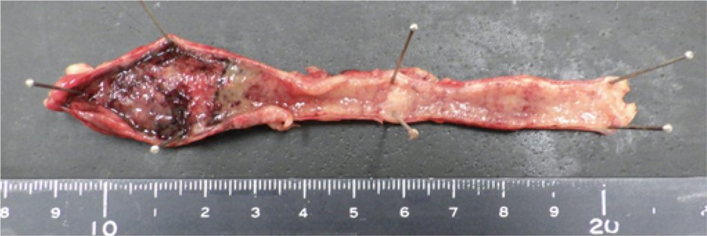
Figure 1
Macroscopic findings of removed appendix vermiformis. Dark colored mucosa was seen in the tip (left of the image).
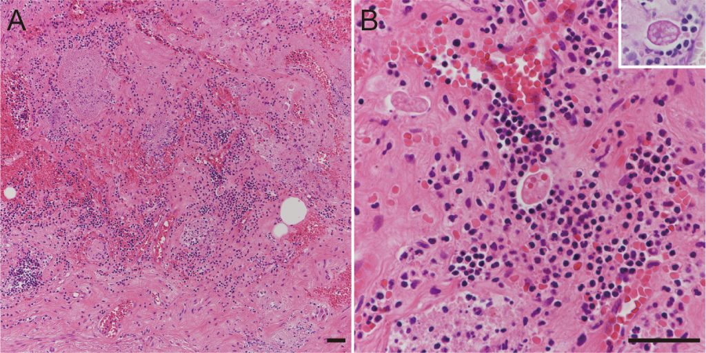
Figure 2 Histopathological features of amebic appendicitis.
A. Moderate-power view showing scattered amebic trophozoites within appendiceal submucosa.
B. High-power view of oval amebic trophozoites with foamy cytoplasm and eosinophilic nucleus-like structures. Periodic acid-Schiff stain highlighting reddish features of trophozoite (inset) (A, hematoxylin and eosin stain, ×100; B, hematoxylin and eosin stain, ×400; inset, periodic acid-Schiff stain, ×400, black bars, 50 μm).
IV. Discussion
E. histolytica infection, or amebiasis, is the second leading cause of death from parasitic infection worldwide.6) In Japan, the annual incidence of amebiasis is low (approximately 500 cases per year) owing to a clean water supply,7) and amebic appendicitis is extremely rare. Our review of Japanese literature revealed only 18 previous reports describing surgically treated amebic appendicitis.5),8) There is a male preponderance in the Japanese incidence of amebic infections, including colitis, liver abscess, and appendicitis.7) In Japan, amebiasis is typically found in men who have sex with men and in individuals with recent travel to endemic areas. However, the present case had no history of travel abroad, and the infectious route was unclear.
Amebic colitis is not uncommon and is usually suspected based on clinical symptoms of bloody and/or mucous diarrhea and is diagnosed by colonoscopic, serological, or stool examination.1) However, amebic appendicitis usually does not show bloody and/or mucous stools suggestive of amebic colitis, although appendicitis sometimes accompanies ileocecal amebic colitis.3),4) The clinical symptoms of previously reported cases of amebic appendicitis and the present case were similar to those of non-amebic appendicitis, including right lower quadrant pain, fever, abdominal tenderness, and guarding. Therefore, the preoperative suspicion of amebic infection is considered difficult. Furthermore, in the present case, blood eosinophilia was not observed despite parasitic infection. Our review of the literature revealed four previous articles on amebic colitis or appendicitis describing the presence or absence of eosinophilia (Table 1). Three of these articles described three cases of amebic colitis without blood eosinophilia.9)-11) The other article investigated amebic appendicitis in the pediatric population and revealed blood eosinophilia in only 14.2% of cases.12) Therefore, blood eosinophilia is not always observed in patients with amebic appendicitis or colitis.
Previous systematic reviews of worldwide amebic appendicitis showed a mortality rate of 3.3%,3) higher than that of overall acute appendicitis (0.09–0.24%),13) although almost half of the cases included in this review were from endemic countries where metronidazole (medication for amebiasis) is routinely administered to all cases of acute appendicitis.14) The mortality rate of amebic appendicitis is reported to be higher in Japan (25%).5) This higher mortality rate would be attributed not only to post-operative complications such as amebic pancolitis, colonic perforation, peritonitis but also to delay for specific treatments.4),8) Discrimination between amebic and non-specific appendicitis should be required.
The diagnosis of amebic appendicitis may be challenging not only clinically but also pathologically. In previous cases,4),5) the first pathological examination of appendectomized specimens failed to reveal amoebas, and pathological reexamination based on clinical indications led to the accurate diagnosis of amebic appendicitis. In the present case, the amebic trophozoites involved not only the surface but also the appendiceal wall. Similar amebic distribution has been reported previously 15),16) and seemed to differ from the surface-predominant lesions of amebic colitis.17) The appendiceal muscularis propria is rich in ganglion cells that mimic amebic trophozoites, which may contribute to their being overlooked at first glance. Furthermore, one pediatric study of amebic appendicitis reported higher perforation rate in wall involvement cases than in luminal colonization cases.12) Therefore, pathologists should be aware of the characteristic features of amebic appendicitis.
In amebic appendicitis, early metronidazole treatment lowers the mortality rate.3) Paromomycin, after metronidazole, is recommended to eliminate intestinal colonization.1) In the present case, an amebic infection predominantly involving the deep appendiceal wall may have been related to resistance to metronidazole treatment and relapse.
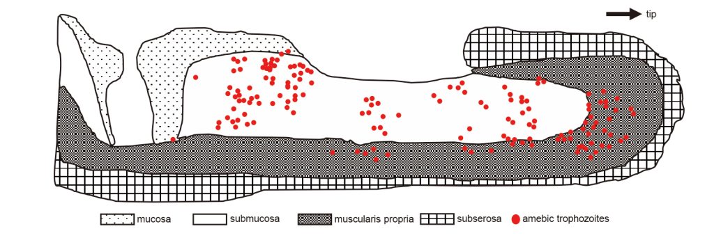
Figure 3 Schematic distribution of amebic trophozoites within longitudinal section of the vermiform appendix. Amebic trophozoites predominantly involving the submucosa and muscularis propria, rather than appendiceal surface.
Table 1 Relationship between blood eosinophilia and intestinal amebiasis.

M, male; F, female; AA, amebic appendicitis; IA, acute appendicitis with incidental amebiasis (amebae found only within the appendiceal lumen)* The total number of cases showing blood eosinophilia was not available from the article.
V. Conclusion
This case report describes an unusual case of acute appendicitis. In this case, eosinophilia was not observed, and E. histolytica trophozoites, highlighted by PAS staining, diffusely involved the removed vermiform appendiceal wall. Physicians should be aware of the presence of amebic appendicitis mimicking non-amebic appendicitis.
Author contribution
Kurihara A contributed to research and writing. Tadakazu A contributed to the editing of clinical findings. Takeo H and Matsukuma S contributed to the editing of pathological and laboratory findings.
Conflict of interest: The authors declare no conflicts of interest.
Funding: None.
Acknowledgements: We would like to thank Editage (www.editage.jp) for English language editing.
References
- Haque R, Huston CD, Hughes M, et al. Amebiasis. N Engl J Med 2003; 348(16): 1565-73. doi: 10.1056/NEJMra0227102.
- Horiki N, Furukawa K, Kitade T, et al. Endoscopic findings and lesion distribution in amebic colitis. J Infect Chemother. 2015; 21(6): 444-8. doi: 10.1016/j.jiac.2015.02.004.PubMed
- Otan E, Akbulut S, Kayaalp C. Amebic acute appendicitis: systematic review of 174 cases. World J Surg 2013; 37(9): 2061-73. doi: 10.1007/s00268-013-2079-5.PubMed
- Suzuki Y, Adachi Y, Yasumizu R, et al. [Amebiases: Two autopsy cases where diagnosis could not be established during the lifetime of the patients] Seizen ni shindan no konnan de atta sekiri ameba kansensyo no 2 boukenrei. Jpn J Diagn Pathol. 2005; 22(1): 25-8 (in Japanese).
- Ito D, Hata S, Shimizu S, et al. Amebiasis presenting as acute appendicitis: Report of a case and review of Japanese literature. Int J Surg Case Rep 2014; 5(12): 1054-7. doi: 10.1016/j.ijscr.2014.10.035.PubMed
- Kantor M, Abrantes A, Estevez A, et al. Entamoeba histolytica: Updates in clinical manifestation, pathogenesis, and vaccine development. Can J Gastroenterol Hepatol 2018; 2018: 4601420. doi: 10.1155/2018/4601420.PubMed
- Ishikane M, Arima Y, Kanayama A, et al. Epidemiology of domestically acquired amebiasis in Japan, 2000−2013. Am J Trop Med Hyg 2016; 94(5): 1008-14. doi: 10.4269/ajtmh.15-0560.
- Sugiyama Y, Osaka Y, Kato F, et al. A case of amebic appendicitis with abdominal wall necrosis, intestinal perforation, and infectious myocarditis after surgery. J Jpn Surg Assoc. 2021; 82(8): 1537-42. doi: 10.3919/jjsa.82.1537 (in Japanese).
- Shijubou N, Sumi T, Kamada K, et al. Fulminant amebic colitis in a patient with concomitant cytomegalovirus infection after systemic steroid therapy: A case report. World J Clin Cases 2021; 9(15): 3726-32. doi: 10.12998/wjcc.v9.i15.3726.PubMed
- Ohnishi K, Murata M, Okuzawa E. Symptomatic amebic colitis in a Japanese homosexual AIDS patient. Intern Med 1994; 33(2): 120-2. doi: 10.2169/internalmedicine.33.120.PubMed
- Yasumura K, Aoyama I, Shibata K, et al. [Early endoscopic diagnosis can save lives and reduce the extent of colon resection for acute fulminant amebic colitis: a case report with review]. Nihon Shokakibyo Gakkai Zasshi. 2022; 119(6): 540-50. doi: 10.11405/nisshoshi.119.540 (in Japanese). PubMed
- Echeverry JME, Valero JJ, Jaramillo LE, et al. Clinical features of amoebic appendicitis in children: A study of 23 cases. J Pediatr Surg 2021; 56(8): 1362-4. doi: 10.1016/j.jpedsurg.2020.12.027.PubMed
- Bhangu A, Søreide K, Saverio SD, et al. Acute appendicitis: modern understanding of pathogenesis, diagnosis, and management. Lancet 2015; 386(10000): 1278-87. doi: 10.1016/S0140-6736(15) 00275-5.PubMed
- Guzmán-Valdivia G. Acute amebic appendicitis. World J Surg 2006; 30(6): 1038-42. doi: 10.1007/s00268-005-0104-z.
- Goyal A, Goyal S, Gupta CR. Acute amoebic appendicitis: An unusual presentation of a usual infection. Indian J Pathol Microbiol. 2019; 62(1): 169-70. doi: 10.4103/IJPM.IJPM_576_17.PubMed
- Kleitsch WP, Kisner P. Amebic appendicitis, peritonitis and wound infection. Ann Surg 1951; 133(1): 139-42. doi: 10.1097/00000658-195101000-00017.PubMed
- Yue B, Meng Y, Zhou Y, et al. Characteristics of endoscopic and pathological findings of amebic colitis. BMC Gastroenterol. 2021; 21(1): 367. doi: 10.1186/s12876-021-01941-z.PubMed