†Hiroko Tsuda, Kenta Noguchi, Mana Miya Kawae*
Tsuda H, Noguchi K, Kawae MM. Influence of high-fat and high-carbohydrate meals on lipids, apolipoproteins, and coagulation and anticoagulation factors. Lab Med Int 2024; 3(1): 15-21. do: 10.51041/lmi.3.1_15
Original
Lab Med Int 2024; 3(1): 15-21
†Correspondence: Department of Nutritional Sciences, Faculty of Nutritional Sciences, Nakamura Gakuen University, 5-7-1 Befu, Jonan-ku, Fukuoka 814-0198, Japan.
E-mail: tsuda”@”nakamura-u.ac.jp
Received December 5, 2023; accepted January 30, 2024
Department of Nutritional Sciences, Faculty of Nutritional Sciences, Nakamura Gakuen University, Fukuoka, Japan.
ABSTRACT
Background: Nonfasting triglyceride levels have an equal or greater impact on predicting cardiovascular disease events than fasting levels.
Objectives: To examine the effects of consuming a high-fat or a high-carbohydrate meal on the plasma levels of lipids, apolipoproteins, and coagulation and anticoagulation factors.
Methods: In a randomized cross-over study, 12 young non-obese women were served isocaloric meals, predominantly fat (69 E% fat) or carbohydrate (91 E% carbohydrates), on two separate days, and blood samples were taken before and 2.5 and 5.5 h after the meal.
Results: After consuming a high-fat meal, there were significant increases in triglycerides, small dense LDL (sdLDL) cholesterol, and factor VII activity. However, levels of total, LDL, and HDL cholesterol, and apolipoproteins decreased; and levels of total protein S antigen and its activity, free protein S antigen, protein C antigen, and fibrinogen remained unchanged. On the other hand, all these parameters, except for apoC-II and total protein S antigen, decreased after eating a high-carbohydrate meal. Insulin increased postprandially, while glucose levels remained unchanged. Strong positive relationships between triglycerides and the levels of sdLDL cholesterol and apoC-II were observed both at fasting and after consuming meals. However, no correlation was found between triglycerides and coagulation and anticoagulation factors such as factor VII activity and total protein S antigen and its activity.
Conclusions: Consuming a high-fat meal increased factor VII activity during the postprandial elevation of triglycerides and sdLDL cholesterol. However, the activated protein C-dependent anticoagulant system may not regulate this postprandial hypercoagulable state.
〔Lab Med Int 2024; 3(1): 15-21〕
Key Words
High-fat or high-carbohydrate meal, triglycerides, factor VII, protein S, small dense LDL cholesterol
I. Introduction
Plasma lipid profiles, including lipids, lipoproteins, and apolipoproteins, are affected by food intake and show a 24-h cycle. Therefore, they are evaluated in blood drawn after fasting for at least 8 h, which normally only occurs a few hours before breakfast 1). Elevated low-density lipoprotein (LDL) cholesterol is a well-established independent risk factor for coronary heart disease (CHD) 2); however, the importance of elevated triglycerides remains to be elucidated. Triglyceride-rich lipoproteins and their remnant lipoproteins are increased in plasma postprandially and may be atherogenic 3) 4). Prospective cohort studies have reported that elevated levels of nonfasting triglycerides have an equal or greater impact on predicting cardiovascular disease (CVD) events and mortality than fasting levels 5)-9).
Postprandial triglyderidemia, particularly after eating high-fat meals, leads to increased levels of plasma factor VII activity and activated factor VII 10). In vitro, large very-low-density lipoproteins (VLDL) rather than small VLDL enhance tissue factor (TF)-independent activation of factor VII by factor Xa and factor Xa/Va 11). In addition, vitamin K-dependent coagulation and anticoagulation factors, such as factors VII, IX, X, prothrombin, protein C, and protein S, as well as C4b-binding protein (C4BP), a complement regulatory protein, have been reported to be associated with triglyceride-rich lipoproteins, chylomicrons, chylomicron remnants, and VLDL, especially after consuming high saturated fat meals; however, there is no association with LDL or high-density lipoprotein (HDL) 12) 13).
Protein S plays an important role in anticoagulation as a cofactor for the activated protein C (APC)-dependent inactivation of factors Va and VIIIa. It also functions as a cofactor for the tissue factor pathway inhibitor
(TFPI)-dependent inactivation of factors Xa and TF-VIIa complex 14) 15). In human plasma, approximately 40% of protein S circulates in its free form, with the remaining 60% of protein S non-covalently binding to C4BP and losing its cofactor activities. Using a cross-sectional study, we previously reported that apolipoprotein (apo) C-II, a critical cofactor of lipoprotein lipase 16), is a significant predictor of fasting total protein S antigen levels in middle-aged obese women, about half of whom exhibited dyslipidemia, as well as in nonobese young women with normolipidemic profiles 17).
Plasma LDL comprises multiple distinct subspecies and has been grouped into four major subclasses based on density, LDL-I through -IV, from the largest, most buoyant to the smallest, most dense 18). These subspecies differ in their metabolic behavior and pathogenic roles. An elevated level of small dense LDL (sdLDL) particles is accepted as an emerging risk factor for CHD 2) 19). They are largely formed from the delipidation of triglyceride-rich VLDL catalyzed by lipoprotein lipase and hepatic lipase 18). Recently, an automated homogeneous assay was developed that accurately measures sdLDL cholesterol 20). A systematic review and meta-analysis reported a positive association between fasting level of sdLDL cholesterol and CHD 21).
The aim of this study was to investigate the impact of consuming a high-fat meal on the levels of lipids and apolipoproteins, including triglycerides, sdLDL cholesterol, and apo C-II, as well as coagulation and anticoagulation factors such as factor VII and protein S, and to compare the findings with those of an isoenergic high-carbohydrates (low-fat) meal used as a control.
II. Methods
Study subjects and study design
Thirteen healthy Japanese female university students were enrolled in this study. The students were not current smokers, did not have menstruation disorders, nor use oral contraceptives. Gene analysis showed that all subjects were homozygous for the wild-type of protein S Tokushima (p.Lys196Glu, rs121918474), a protein S gene polymorphism prevalent among the Japanese population 22). One subject dropped out of the study due to feeling sick while consuming a test meal. The basic characteristics (mean ± SD) of the final 12 participants were as follows: age, 21.3 ± 0.5 years; height, 160.8 ± 5.6 cm; weight, 56.6 ± 6.5 kg; BMI, 21.9 ± 2.2 kg/m2. The present study was performed after approval of the Ethics Committee of Nakamura Gakuen University and written informed consent was obtained from all participants.
The trial was a cross-over study with the order of the meals randomized. The day before each study, the participants were directed to refrain from eating high-fat meals, drinking alcohol, and carrying out intense activity. On the day of the study, all participants consumed an experimental isoenergic high-fat or high-carbohydrate meal after an overnight fast. The meals were served on two separate days (seven to twenty days apart; average: 11 days) at admission to the Health Promotion Center of Nakamura Gakuen University. After measuring weight and height and taking a blood sample, the participants consumed test meals with water under observation between 8:45 and 9:00 in the morning. High-fat meals (total energy 705 kcal: 69 E% fat, 27 E% carbohydrate, and 4 E% protein) consisting of bread, butter, margarine, and pudding, and high-carbohydrate meals (total energy 701 kcal: 2 E% fat, 91 E% carbohydrate, and 5 E% protein) consisting of rice and nonfat agar jelly were consumed. Then the participants were allowed to rest in a sitting position and were instructed not to consume anything except for water (<500 mL during the study). Blood samples were drawn at 2.5 and 5.5 h after the meal based on a standardized oral fat tolerance test 10), with a slight modification.
Analyses of blood samples
Plasma and serum samples were prepared and stored as reported previously 17). The analysis of the antigen level and APC-cofactor activity of total protein S was performed by using a total protein S-assay system (Shino-Test Co., Sagamihara, Japan) 23). Free protein S and protein C antigen were measured using latex agglutination methods (Diagnostica Stago, Inc., New Jersey, USA and LSI Medience. Co., Tokyo, Japan, respectively), factor VII activity was by a clotting assay (Instrumentation Laboratory Co., Bedford, USA), and fibrinogen by a thrombin-time method (Sysmex Co., Kobe, Japan). Total, LDL, and HDL cholesterol, triglycerides, apoA-I, B (the sum of B-48 and B-100), C-II, C-III, E, glucose, and insulin were measured using previously reported methods 17). The level of sdLDL cholesterol was assayed by an automated homogeneous assay (Denka Seiken Co., Tokyo, Japan) 20). Genomic DNA was purified from the buffy coat of citrated blood samples, and the genotype of protein S Tokushima mutation was assessed, as reported previously 23).
Statistical analysis
Kolmogorov-Smirnov test was used to test the normality of data distribution, and all data were found to be normally distributed. Paired Student’s t test was used to evaluate the fasting levels of all measures on two different study visits and there was no significant difference. A one-way repeated measures ANOVA followed by multiple comparisons was used to test for differences across time. We performed univariate linear regression analyses to determine whether there were any correlations between fasting values (averaged of two visits), postprandial values, and changes in values during blood sampling, and Pearson’s correlation coefficients were calculated. Statistical analyses were performed using PASW Statistics ver. 18 (SPSS Inc., Chicago, IL, USA), and a P value < 0.05 was considered significant.
III. Results
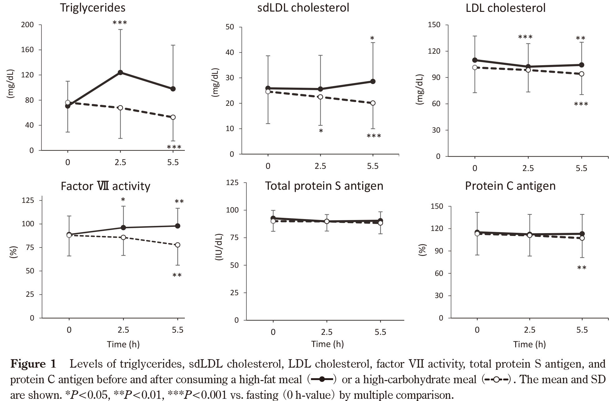
After consuming a high-fat meal, triglycerides significantly increased at 2.5 h and decreased afterward (Table 1, Figure 1). The level of sdLDL cholesterol increased after 5.5 h; however, total, LDL, and HDL cholesterol, as well as apo A-I, B, C-II, C-III, and E decreased postprandially. Accordingly, the ratio of sdLDL cholesterol to LDL cholesterol showed a significant increase 5.5 h after the meal. A significant increase in factor VII activity was found over time after the meal, whereas the levels of fibrinogen, total PS antigen and its activity, free protein S antigen, and protein C antigen remained unchanged. Insulin levels peaked 2.5 h after meal consumption, while glucose levels remained unchanged. On the other hand, after consuming a high-carbohydrate meal, all the levels of lipids, apolipoproteins, and coagulation and anticoagulation factors decreased, except for apo C-II and total protein S antigen and its activity. Apo C-II increased 2.5 h after the meal, while total protein S antigen and its activity remained unchanged. Insulin peaked 2.5 h after the meal and the peak was higher than after consuming a high-fat meal, but glucose remained unchanged. The postprandial decrease in total, LDL, HDL cholesterol, and most apolipoprotein may be due to hemodilution 24), because the participants consumed approximately 500 mL of water during the study.
We then conducted univariate linear regression analyses to identify the factors associated with triglycerides (Table 2). During fasting, there were strong positive relationships between triglycerides and the levels of sdLDL cholesterol, sdLDL cholesterol/LDL cholesterol ratio, and apo C-II, and these relationships became stronger after consuming a high-fat or high-carbohydrate meal. In contrast, triglyceride levels were positively correlated with total and LDL cholesterol only after consuming a high-fat meal, and with apoB and apo C-III after consuming both high-fat and high-carbohydrate meals. No correlation was found between triglycerides and any other variables, including coagulation and anticoagulation factors such as factor VII activity, protein S antigen and its activity, and protein C antigen in both fasting and postprandial states. Furthermore, correlation analyses did not reveal any statistically significant associations between factor VII activity and the levels of triglycerides or sdLDL cholesterol (data not shown), nor between the postprandial changes in factor VII activity and those in triglycerides and sdLDL cholesterol (Table 3).
Table 1 Fasting and postprandial levels of lipids, apolipoproteins, and coagulation and anticoagulation factors.
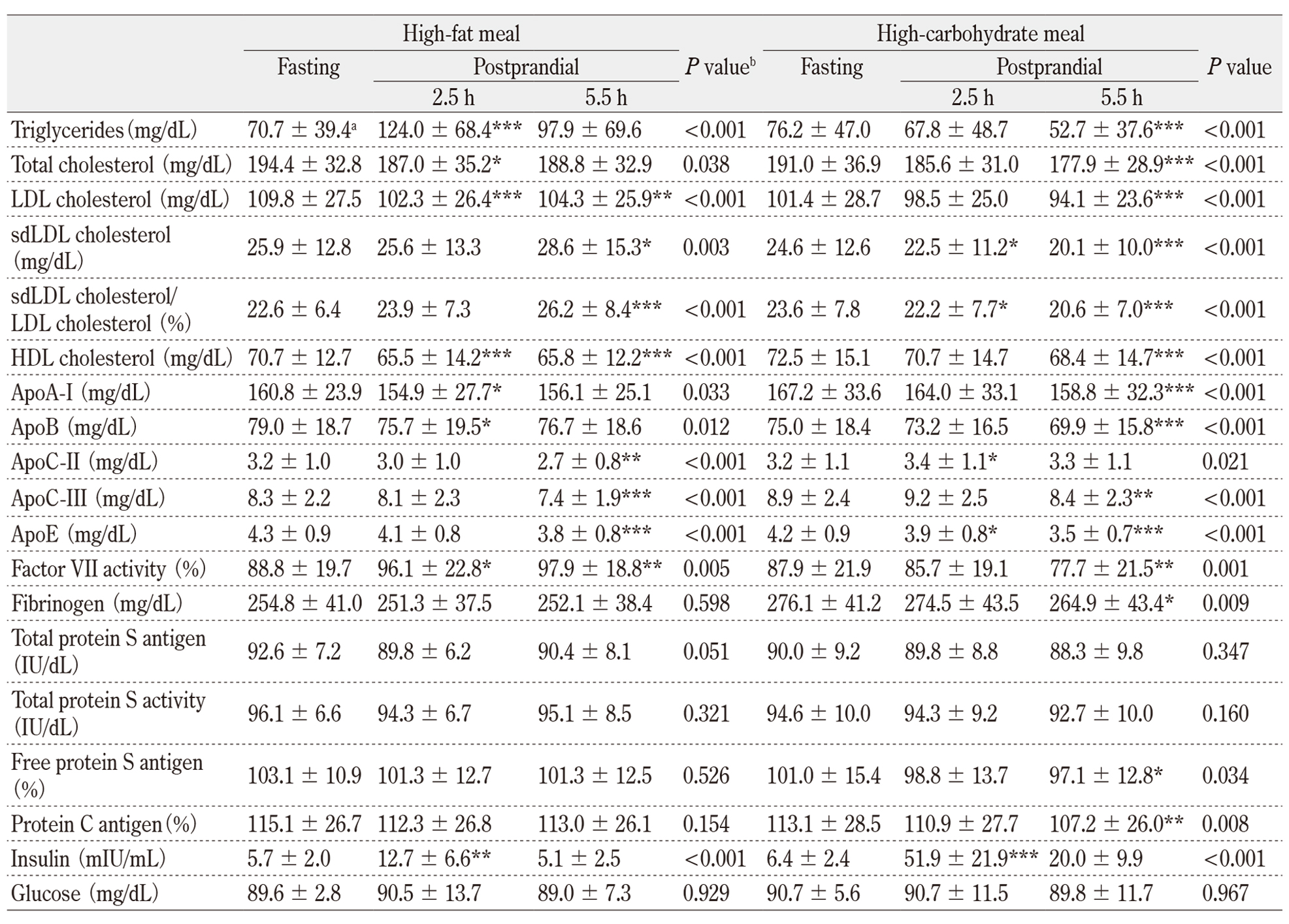
aMean ± SD, bone-way repeated measures ANOVA to test for differences across time. *P<0.05, **P<0.01, ***P<0.001 vs. fasting by multiple comparisons.
Abbreviations, LDL: low-density lipoprotein, sdLDL, small dense LDL; HDL, high-density lipoprotein; Apo, apolipoprotein.
IV. Discussion
This study demonstrated that consuming a high-fat meal but not a high-carbohydrate (low-fat) meal increased factor VII activity during the postprandial elevation of triglycerides and sdLDL cholesterol. However, total protein S antigen and its APC-cofactor activity, free protein S antigen, and protein C antigen levels did not change postprandially. Strong positive relationships were observed between triglycerides and the levels of sdLDL cholesterol and apoC-II both at fasting and after consuming meals.
Activated factor VII (VIIa) and VIIa-antithrombin complex are reported to significantly increase after eating high-fat meals, indicating postprandial activation of factor VII 10) 25) 26). An in vitro study using purified systems reported that large VLDL, rather than small VLDL, enhances TF-independent activation of factor VII by factor Xa and factor Xa/Va 11). However, we did not find any significant associations between factor VII activity and the levels of triglycerides or sdLDL cholesterol, nor between changes in factor VII activity and changes in the levels of triglycerides and sdLDL cholesterol during food consumption. Some in vivo studies have shown that factor IX 10) or kallikrein 27) is essential for the postprandial activation of factor VII; however, the detailed mechanisms are not yet understood.
After consuming high saturated fat meals, protein S, protein C, and C4BP as well as vitamin K-dependent coagulation factors such as factors VII have been reported to be associated with triglyceride-rich lipoproteins, chylomicrons, chylomicron remnants, and VLDL 12) 13). However, our study found no significant changes in the levels of total protein S antigen and its APC-cofactor activity, free protein S antigen, and protein C antigen levels after a high-fat meal. Protein S plays a critical role in anticoagulation as a cofactor for the APC-dependent and TFPI-dependent pathways 14) 15). Previous research has shown that the consumption of high-fat meals has no effect on plasma levels of free TFPI 26) 27). Taken together, the APC-dependent and TFPI-dependent anticoagulation pathways may not regulate the postprandial activation of factor VII after consuming high-fat meals.
Our earlier report demonstrated that apoC-II, a critical cofactor of lipoprotein lipase 16), is a significant predictor of fasting total protein S antigen levels 17). In the present study, we observed a weak correlation (r = 0.522, P = 0.082) between total protein S antigen and apoC-II during fasting; however, this correlation disappeared after consuming meals (data not shown). We also found that of all the apolipoproteins, only apoC-II had a significant correlation with triglycerides, both during fasting and after consuming meals. This may explain why apoC-II has become a potential target for developing new drugs to treat hypertriglyceridemia 16).
In line with a previous report 27), we observed that insulin levels were significantly higher after eating a high-carbohydrate meal compared to a high-fat meal; however, factor VII activity decreased after eating a high-carbohydrate meal, suggesting that there was little or no factor VII activation. A study conducted on healthy individuals who were exposed to 24 h of hyperinsulinemia with or without hyperglycemia reported that factor VII activity decreased the most during high glucose/high insulin and to a lesser degree during selective hyperinsulinemia and selective hyperglycemia 28).
Ogita et al. 29) have reported that sdLDL cholesterol of young adults remains unchanged for up to 4 h after consuming a fat-rich test material (17 g fat/m2 of body surface area), while triglycerides peak at 2 h after the meal. In our study, the amount of fat consumed was an average of 35 fat/m2 of body surface area, which was twice as much as their study. As a result, consuming meals containing a high amount of fat tends to raise the levels of sdLDL cholesterol. The homogeneous assay of sdLDL cholesterol measures cholesterol in LDL particles with a density of 1.044-1.63 g/mL 20), which corresponds with very small LDL-IV 18). The sdLDL particles are formed from the lipolysis of large triglyceride-rich VLDL by lipoprotein lipase and hepatic lipase 18). This may explain why the increase in sdLDL cholesterol was observed 3 h later than the increase in triglycerides after consuming a high-fat meal. As expected, significant correlations were found between triglycerides and sdLDL cholesterol during fasting and after consuming a high-fat or a high-carbohydrate meal. The atherogenic potential of sdLDL includes prolonged plasma residence, increased susceptibility to oxidative modification, and a greater tendency to penetrate into the arterial wall 18) 30). Therefore, the postprandial elevation of sdLDL cholesterol may contribute to the relationship between elevated levels of nonfasting triglycerides and the prediction of CVD events and mortality 5)-9).
The present study had several limitations. First, the study had a small sample size, which may reduce the ability to detect significant changes from fasting to nonfasting conditions and postprandial changes. Second, the study only included young women as subjects. We previously found that the relationship between protein S and apoC-II is much stronger in middle-aged obese women, about half of whom exhibited dyslipidemia, than in young nonobese women 17). Third, we analyzed only factor VII and fibrinogen as coagulation factors; however, previous studies reported that prothrombin, factors VII, IX, X are associated with triglyceride-rich lipoproteins in both fasting and postprandial plasma 12). Fourth, we did not analyze TFPI-cofactor activity of protein S.
Table 2 Relationships between triglycerides and the levels of other lipids, apolipoproteins, and coagulation and anticoagulation factors.
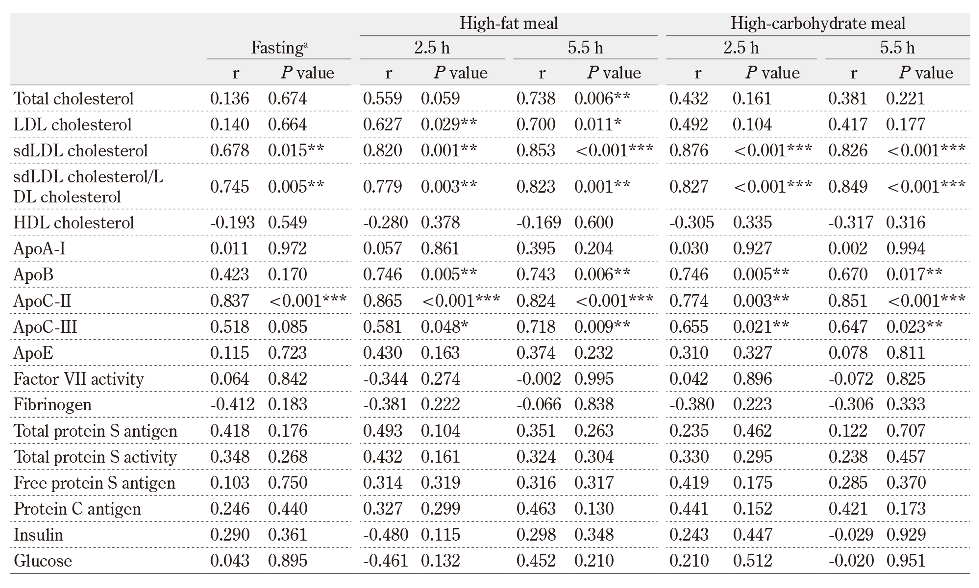
aFasting levels of all measures were the means of the data obtained at two visits. r, Pearson’s correlation coefficient: *P<0.05, **P<0.01, ***P<0.001
Details of the abbreviations used are shown in Table 1.
Table 3 Relationships between changes of factor VII activity and those of triglycerides and sdLDL cholesterol during food intakes

FVII:C, factor VII activity; △2.5-0, changes between fasting and 2.5 h postprandial: △5.5-2.5, changes between 2.5 h postprandial and 5.5 h postprandial: sdLDL, small dense LDL; r, Pearson’s correlation coefficient.
Acknowledgments
This study was supported by JSPS KAKENHI Grant Number JP18K07486. We thank Dr. Tomohide Tsuda and Dr. Xiuri Jin of Shinotest Corporation for measuring total protein S antigen and its activity.
Authorship Contributions
H. Tsuda and K. Noguchi designed the research. H. Tsuda, K. Noguchi, and M. M. Kawae performed the study and analyzed the data. H. Tsuda wrote the manuscript. All authors read and approved the final version of the paper.
Disclosure of Conflicts of Interest
The authors declare that they have no conflict of interest.
References
- Nordestgaard BG. A test in context: Lipid profile, fasting versus nonfasting. J Am Coll Cardiol 2017; 70(13): 1637-46.PubMed
- National Cholesterol Education Program Expert Panel on Detection, Evaluation, Treatment of High Blood Cholesterol in Adults. Third Report of the National Cholesterol Education Program (NCEP) Expert Panel on Detection, Evaluation, and Treatment of High Blood Cholesterol in Adults (Adult Treatment Panel III) final report. Circulation. 2002; 106(25): 3143-421.PubMed
- Zilversmit DB. Atherogenic nature of triglycerides, postprandial lipidemia, and triglyceride-rich remnant lipoproteins. Clin Chem 1995; 41(1): 153-8.PubMed
- Ginsberg HN. New perspectives on atherogenesis: role of abnormal triglyceride-rich lipoprotein metabolism. Circulation 2002; 106(16): 2137-42.PubMed
- Eberly LE, Stamler J, Neaton JD, et al. Relation of triglyceride levels, fasting and nonfasting, to fatal and nonfatal coronary heart disease. Arch Intern Med 2003; 163(9): 1077-83.PubMed
- Bansal S, Buring JE, Rifai N, et al. Fasting compared with nonfasting triglycerides and risk of cardiovascular events in women. JAMA 2007; 298(3): 309-16.PubMed
- Nordestgaard BG, Benn M, Schnohr P, et al. Nonfasting triglycerides and risk of myocardial infarction, ischemic heart disease, and death in men and women. JAMA 2007; 298(3): 299-308.PubMed
- Iso H, Imano H, Yamagishi K, et al. Fasting and non-fasting triglycerides and risk of ischemic cardiovascular disease in Japanese men and women: the Circulatory Risk in Communities Study (CIRCS). Atherosclerosis 2014; 237(1): 361-8.PubMed
- Hirata A, Okamura T, Hirata T, et al. Relationship between non-fasting triglycerides and cardiovascular disease mortality in a 20-year follow-up study of a Japanese general population: NIPPON DATA90. J Epidemiol 2022; 32(7): 303-13.PubMed
- Silveira A. Postprandial triglycerides and blood coagulation. Exp Clin Endocrinol Diabetes 2001; 109(4): S527-32.PubMed
- Kjalke M, Silveira A, Hamsten A, et al. Plasma lipoproteins enhance tissue factor-independent factor VII activation. Arterioscler Thromb Vasc Biol 2000; 20(7): 1835-41.PubMed
- Xu N, Dahlbäck B, Ohlin AK, et al. Association of vitamin K-dependent coagulation proteins and C4b binding protein with triglyceride-rich lipoproteins of human plasma. Arterioscler Thromb Vasc Biol. 1998; 18(1): 33-9.PubMed
- Zhou L, Xu N, Nilsson A. Association of prothrombin and protein S with plasma triglyceride-rich lipoproteins in humans after test meals rich in milk fat or soybean oil. Thromb Res 2002; 108(4): 209-13.PubMed
- Dahlbäck B. Vitamin K-dependent protein S: Beyond the protein C pathway. Semin Thromb Hemost 2018; 44(2): 176-84.PubMed
- Gierula M, Ahnström J. Anticoagulant protein S-New insights on interactions and functions. J Throm Haemost 2020; 18(11): 2801-11.PubMed
- Wolska A, Reimund M, Remaley AT. Apolipoprotein C-II: the re-emergence of a forgotten factor. Curr Opin Lipidol 2020; 31(3): 147-53.PubMed
- Otsuka Y, Ueda M, Nakazono E, et al. Relationship between plasma protein S levels and apolipoprotein C-II in Japanese middle-aged obese women and young nonobese women. Blood Coagul Fibrinolysis 2018; 29(1): 39-47.PubMed
- Berneis KK, Krauss RM. Metabolic origins and clinical significance of LDL heterogeneity. J Lipid Res 2002; 43(9): 1363-79.PubMed
- Boren J, Chapman MJ, Krauss RM, et al. Low-density lipoproteins cause atherosclerotic cardiovascular disease: pathophysiological, genetic, and therapeutic insights: a consensus statement from the European Atherosclerosis Society Consensus Panel. Eur Heart J 2020; 41(24): 2313-30.PubMed
- Ito Y, Fujimura M, Ohta M, et al. Development of a homogeneous assay for measurement of small dense LDL cholesterol. Clin Chem 2011; 57(1): 57-65.PubMed
- Liou L, Kaptoge S. Association of small, dense LDL-cholesterol concentration and lipoprotein particle characteristics with coronary heart disease: A systematic review and meta-analysis. PLoS One 2020; 15(11): e0241993.PubMed
- Tsuda H, Noguchi K, Oh D, et al. Racial differences in protein S Tokushima and two protein C variants as genetic risk factors for venous thromboembolism. Res Pract Thromb Haemost 2020; 4(8): 1295-300.PubMed
- Noguchi K, Nakazono E, Tsuda T, et al. Plasma phenotypes of protein S Lys196Glu and protein C Lys193del variants prevalent among young Japanese women. Blood Coagul Fibrinolysis 2019; 30(8): 393-400.PubMed
- Langsted A, Freiberg JJ, Nordestgaard BG. Fasting and nonfasting lipid levels: influence of normal food intake on lipids, lipoproteins, apolipoproteins, and cardiovascular risk prediction. Circulation 2008; 118(20): 2047-56.PubMed
- Silveira A, Carlo A, Adam M, et al. VIIaAT complexes, procoagulant phospholipids, and thrombin generation during postprandial lipemia. Int J Lab Hematol 2018; 40: 251-7.PubMed
- Landgrebe LE, Juhl CB, Andersen V, et al. Postprandial factor VII activation does not increase plasma concentrations of prothrombin fragment 1 + 2 in patients with morbid obesity. Thromb Res 2020; 196: 260-7.PubMed
- Larsen LF, Marckmann P, Bladbjerg EM, et al. The link between high-fat meals and postprandial activation of blood coagulation factor VII possibly involves kallikrein. Scand J Clin Lab Invest 2000; 60(1): 45-54.PubMed
- Vaidyula VR, Rao AK, Mozzoli M, et al. Effects of hyperglycemia and hyperinsulinemia on circulating tissue factor procoagulant activity and platelet CD40 ligand. Diabetes 2006; 55(1): 202-8.PubMed
- Ogita K, Ai M, Tanaka A, et al. Serum concentration of small dense low-density lipoprotein-cholesterol during oral glucose tolerance test and oral fat tolerance test. Clin Chim Acta 2008; 387(1-2): 36-41.PubMed
- Krauss RM. Small dense low-density lipoprotein particles: clinically relevant? Curr Opin Lipidol 2022; 33(3): 160-6.PubMed