Jeong Hui Kim*1, Ryosuke Kikuchi*2†, Atsuo Suzuki*1, Rika Watarai*1, Kaori Goto*1, Yui Okumura*1, Tadashi Matsushita*3, 4
Kim JH, Kikuchi R, Suzuki A, Watarai R, Goto K, Okumura Y, Matsushita T. Comparison of target antigen and immunoglobulin isotypes in anti-SARS-CoV-2 antibodies from natural infection and vaccination. Lab Med Int 2023; 2(3): 60-66. doi: 10.51041/lmi.2.3_60
Original
Lab Med Int 2023; 2(3): 60-66
† Correspondence: Division of Clinical Laboratory, Gifu University Hospital, 1-1 Yanagido, Gifu, 501-1194, Japan.
E-mail: ryosuke”@”gifu-u.ac.jp
Received June 1, 2023; accepted September 7, 2023
*1 Department of Medical Technique, Nagoya University Hospital
*2 Division of Clinical Laboratory, Gifu University Hospital
*3 Department of Transfusion Medicine, Nagoya University Hospital
*4 Department of Clinical Laboratory, Nagoya University Hospital
ABSTRACT
The immune system produces antibodies following SARS-CoV-2 infection and vaccination. However, we lack
comprehensive information about the humoral responses after infection and vaccination, which are similar but might
differ in type or amount of produced antibodies. Therefore, we compared different types and amounts of antibodies
produced by the immune system in response to infection versus those produced by BNT162b2 (Pfizer/BioNTech)
vaccines. Our findings indicated that naturally infected individuals had 14-fold higher anti-SARS-CoV-2 spike protein
(anti-S) and 41-fold higher anti-SARS-CoV-2 receptor-binding domain of the viral spike protein (anti-RBD) IgM
titers than SARS-CoV-2-naïve vaccinees. Remarkably, naturally infected individuals maintained high levels of IgM
antibody titers up to six weeks after symptom onset. A significantly rapid increase in anti-S IgG titers in primary
infection was observed, eventually reaching a level similar to that in people who had third booster vaccination. This
study revealed the characteristics of the target antigen and immunoglobulin isotypes after natural SARS-CoV-2 infection
and COVID-19 vaccination.
〔Lab Med Int 2023; 2(3): 60-66〕
Key Words
SARS-CoV-2, antibody test, CLEIA, natural infection, vaccination
I. Introduction
Coronavirus disease 2019 (COVID-19) has rapidly spread across the globe since its first report in China in December 2019. Owing to their clinical significance, many agents have been investigated as COVID-19 vaccines and evaluated for their efficacy and safety in clinical trials. During the last three years, 183 and 199 COVID-19 vaccine candidates have been developed in clinical and pre-clinical trials, respectively1). The target proteins of SARS-CoV-2 antibodies can be classified into three types: the nucleocapsid protein (anti-N), the spike protein S1 domain (anti-S), and the receptor-binding domain of SARS-CoV-2 spike protein (anti-RBD). Current COVID-19 vaccines principally target the viral S protein or its receptor-binding domain to primarily elicit a robust neutralizing antibody response2). Notably, anti-RBD IgG titers are significantly correlated with the neutralization of authentic SARS-CoV-23)-6). The IgM antibody increases temporarily during the early stages of infection, after which the IgG antibody is produced through a class-switch; this antibody is maintained over a prolonged time7) 8). Therefore, testing for SARS-CoV-2 antibodies could be a valuable approach to assessing the immune response to SARS-CoV-2 infection and the immune response to the vaccination itself.
Immunochromatography and automated immunoassays, such as chemiluminescence immunoassay, electrochemiluminescence immunoassay, and chemiluminescence enzyme immunoassay (CLEIA) are the common SARS-CoV-2 antibody tests routinely used in laboratories. Currently available antibody reagents use the S protein or its receptor-binding domain of SARS-CoV-2 as target antigens, the difference in reactivity against the S or RBD has not been fully revealed. Although several SARS-CoV-2 antibody test kits have been developed as research reagents, they provide insufficient information about their sensitivity and specificity. Furthermore, we have limited information about serum anti-SARS-CoV-2 IgM/IgG levels after both SARS-CoV-2 natural infection and vaccination. The persistence of the effect in preventing infection or the severity of symptoms remains unclarified.
We aimed to clarify the relationship between target antigen and immunoglobulin isotypes by exploring different types and amounts of antibodies conferred by natural infection versus that from the BNT162b2 using six different antibody reagents.
II. Methods
1.Study design and participants Naturally infected individuals
This retrospective cohort study was performed among patients with COVID-19 who were admitted to Nagoya University Hospital (Japan) from April 2020 to September 2020. The study protocol was approved by the Ethics Committee of Nagoya University Hospital (approval number:2020-0095), and we applied an opt-out method to obtain informed consent for participation in this study by using the poster available for every patient in our hospital. All methods were conducted in compliance with the principles of the 1964 Declaration of Helsinki and its later amendments. All 13 unvaccinated hospitalized patients (49 to 81 years old) with primary infections were included in this study. Serum samples were collected from all patients between 0–9 days after symptom onset. The remaining serum samples were collected after routine testing. A total of 197 sequential serum samples collected up to 47 days after symptom onset were analyzed.
2. SARS-CoV-2-naïve individuals
This retrospective cohort study was performed among healthcare workers at Nagoya University Hospital (Japan) from March 2021 to April 2022. The study protocol was approved by the Ethics Committee of Nagoya University Hospital (approval number:2021-0040), and all participants provided written informed consent. All methods were conducted in compliance with the principles of the Declaration of Helsinki and relevant guidelines. All workers who received mRNA BNT162b2 vaccines were invited to participate voluntarily, and 25 SARS-CoV-2-naïve individuals who were going to receive a three-dose regimen of the BNT162b2 vaccine were finally enrolled. Blood was collected at seven time points: day 0 (before the first dose); day 7(seven days after the first dose); day 21(before or up to three days after the second dose); day 91(two months after the second dose); day 183(five months after the second dose); day 274 (before or up to 14 days after the third dose), and day 365. Serum samples were used for antibody testing.
3. Antibody measurements
Anti-SARS-CoV-2 antibody detection was performed using six HISCLTM SARS-CoV-2 antibody reagents (Sysmex, Kobe, Japan) on the HISCLTM-5000 platform9). In this study, the levels of anti-SARS-CoV-2 IgM and IgG antibodies against the nucleocapsid protein (anti-N), spike protein S1 domain (anti-S), and receptor-binding domain (RBD) of SARS-CoV-2 spike protein (anti-RBD) were quantitatively detected. Recombinant S1 and RBD proteins were produced based on the same accession number sequence (YP_009724390). The S1 domain and RBD contained amino acids 12–684 and 319–537, respectively. Recombinant antigens were purified and coupled with magnetic beads as previously described9. In the HISCLTM system, the serum sample was first reacted with magnetic beads coupled with SARS-CoV-2‒ specific recombinant antigens bound to the magnetic beads. After bound/free separation, the antigen-antibody complex was incubated with an alkaline phosphatase-conjugated antibody against human IgG or IgM to form a sandwich immunocomplex. Anti-IgM and -IgG levels were considered reactive or non-reactive, respectively, using cut-off values of 20 and 10 U/mL, respectively.
4. Serum collection
Blood was collected in no additive tubes (plain tubes) and centrifuged at 1,800 g for 10 min. The samples were stored at −80℃ until further antibody analysis.
5. Statistical analysis
All analyses were performed using R statistical software (version 4.2.1; R Foundation for Statistical Computing) and GraphPad Prism software (version 9.3.1, San Diego, CA, USA). Correlation analysis was performed using Spearman’s correlation test. Comparisons between naturally infected and BNT162b2-vaccinated individuals were analyzed using the non-parametric Mann–Whitney U test. For all analyses, statistical significance was indicated as follows: ns (non-significant; p > 0.05), *(p < 0.05), **(p < 0.005), ***(p < 0.0005), ****(p < 0.0001).
III. Results
1. Demographic and baseline characteristics of naturally infected individuals
Thirteen hospitalized patients with COVID-19 with primary infections among unvaccinated individuals were included in the study. The demographic and clinical characteristics are provided in Table 1. The patients had a median age of 73 years (49–81 years), 15.4% were women, and among these, 53.7% were smokers. Fever was the most common symptom (84.6 %) on admission. Cardiovascular disease was the most common comorbidity (46.2 %), followed by hypertension and type 2 diabetes (38.5%). Four patients (30.8%) underwent hemodialysis.
2. Demographic and baseline characteristics of SARS-CoV-2-naïve individuals
Twenty-five SARS-CoV-2-naïve individuals who received a three-dose regimen of the BNT162b2 vaccine were enrolled in this study. The patient characteristics are presented in Table 2. The mean age of the participants was 39 years (23–63 years), and 76.0% were women. The most prevalent self-reported symptoms at the first dose were pain at the injection site (76.0%), myalgia (32.0%), fatigue (12.0%), and pyrexia (8.0%). The most prevalent self-reported symptoms at the second dose were pain at the injection site (60.0%), fatigue (56.0%), myalgia (44.0%), and pyrexia (36.0%). At the third dose, pain at the injection site (64.0%), fatigue (56.0%), pyrexia (52.0%), and chills (36.0%) were observed. The percentage of participants reporting pain at the injection site was stable between the three doses; the occurrence of pyrexia increased from 8.0% (2/25) at the first dose to 52.0% (13/25) at the third dose.
3. SARS-CoV-2 antibody reactivity patterns
We first analyzed the antibody levels produced from the natural infection and that from the BNT162b2 mRNA vaccination. The natural infection resulted in higher anti-nucleocapsid (N) IgM and IgG levels (Figure 1). The median IgG after seven days of symptom onset was 10.6 U/mL (min-max, 0.7–119.5), positive for anti-IgG. In the nucleocapsid, the seroconversion of anti-IgM and IgG occurred simultaneously or sequentially. The BNT162b2 vaccine only encodes the SARS-CoV-2 spike protein, and no anti-N was observed in the vaccinees. Of note, naturally infected individuals maintained high levels of IgM antibody titers up to six weeks after symptom onset. Compared with naturally infected individuals, SARS-CoV-2-naïve vaccines produced a small amount of anti-S and anti-RBD IgM. In the BNT162b2 vaccinees, anti-S IgM and RBD IgM responded only to the first dose and did not respond after the second or third dose.
4. Peak antibody titers
Given that the differences in antibody titers between natural infection and vaccination, we decided to focus on the peak antibody titers in these two groups. In SARS-CoV-2-naïve individuals who had a three-dose regimen of the BNT162b2 vaccine, three peaks were observed; day 21 (before or up to three days after the second dose); day 91 (approximately two months after the second dose); day 274 (before or up to 14 days after the third dose). Anti-S IgM levels peaked three weeks after the first dose, and anti-IgG levels peaked after booster vaccination. (Figure 1) Anti-S IgM and anti-RBD IgM were 14- and 41-fold higher in the naturally infected group than in the vaccinated group, respectively (Figure 2). In the primary infection, a significant rapid increase in IgG titers with anti-S and anti-RBD was noted, similar to that of the second and third booster shots. The anti-RBD IgG titer in participants with three doses of the BNT162b2 vaccine was higher than that in naturally infected patients at a time point after the third vaccine dose was administered (p > 0.0001).
5. Anti-S/RBD ratio
Anti-RBD IgG levels are significantly correlated with the neutralization of authentic SARS-CoV-23)-6). Therefore, we validated the anti-S/RBD ratio at each time point and compared anti-S and anti-RBD between naturally acquired and vaccine-induced immunity. The BNT162b2 vaccine exhibited convergence of the S/RBD IgG ratio with booster vaccination (Figure 3). The S/RBD IgG ratio at 12 months after the first dose was 1.158 (min-max, 0.986–1.490) and the S/RBD IgM ratio was 0.574 (min–max, 0.203–1.151) (Figure 3), suggesting that anti-S IgG reflects anti-RBD IgG changes. In contrast, the natural infection did not result in a constant S/RBD antibody ratio compared with vaccination.
6. Discussion
Several studies have reported a relationship between virus neutralization levels and antibody titers measured using serological tests. Anti-RBD IgG titers significantly correlate with the neutralization of SARS-CoV-23)-6) 10)-13). Therefore, antibody testing can play a critical role in assessing the immune response to SARS-CoV-2 infection and vaccination. To obtain accurate test results, the reactivity of the reagent must be evaluated, and the features and duration of antibody responses to SARS-CoV-2 infection must be defined. Therefore, we evaluated different immunoglobulin isotypes (IgM and IgG) and the levels of antibodies against SARS-CoV-2 N, S, and RBD antigens in longitudinal samples from naturally infected and vaccinated individuals. Our analysis demonstrated that naturally infected individuals maintained high levels of IgM antibody titers up to six weeks after symptom onset, indicating the contribution of antigen-specific memory B cells16)-18). Natural infection could stimulate B cell production via Toll-like receptors (TLRs) to induce downstream signaling, resulting in antibody production and memory B-cell responses16)-18). According to one study, unlike antibodies, virus-specific memory B cells persist at high levels for at least 12 months post-infection15. BNT162b2 mRNA contains N1-methylpseudouridine instead of uridine, which reduces innate immune recognition by TLRs and RIG-I-like receptors19). Vaccination with BNT162b2 mRNA resulted in low IgM antibody titers and low persistence after the first dose, suggesting a primary antibody response by naive B cells20). Taken together, these results suggest that antibody responses during natural infection are maintained by the induction and contribution of IgM memory B cells relative to vaccination.
Naturally infected individuals were diagnosed from April to September 2020, which could be estimated epidemiologically as a mutant strain based on the time of infection21. The BNT162b2 mRNA vaccine is a monovalent vaccine containing the original wild-type spike protein of SARS-CoV-219). As the anti-S and anti-RBD antibodies acquired through infection with mutant strains are derived from antibodies with the mutated S or RBD antigen epitopes, the reactivity of the SARS-CoV-2 antibody reagent may differ from that of wild-type anti-S and anti-RBD antibodies produced by vaccination. Natural infection resulted in higher anti-S and anti-RBD IgG levels, similar to those of participants who had a third booster vaccination; however, caution is warranted in interpreting these data.
In this study, statistical analysis revealed no significant relationship between post-vaccination symptoms and the strength of the antibody responses. However, we observed that most individuals had severe adverse events at the third dose, including pyrexia, and older age was associated with lower titers. We also observed individual differences in the antibody response, and IgM antibody levels helped distinguish people with low and high IgG titers. In the low IgM group, an increase in the IgG titer, comparable to that in the high IgM group, was observed with booster vaccination. This result suggests that higher antibody titers are expected with booster vaccinations, although people have a weakened immune response. Therefore, antibody testing may help generate a vaccination plan for each individual, considering their biochemical background.
Although the S protein or its receptor-binding domain IgG is used to estimate the humoral immune response to the virus, the difference in reactivity against the S or RBD has not been fully revealed. Infectivity-enhancing antibodies recognizing the N-terminal domain (NTD) of the spike are produced following natural infection with SARS-CoV-2. Antibodies to the entire spike protein, including NTD antibodies, may have been produced in the early phase of infection22)23). Thus, the early S/RBD ratio was not as constant as in vaccines. Nevertheless, positive correlation between anti-S IgG and anti-RBD IgG levels was observed not only in the vaccinated group but also in the naturally infected group. Our results are consistent with the results of Wang H et al. 24), who demonstrated that SARS-CoV-2 RBD-IgG were strongly correlated with S-IgG both in severe and non-severe SARS-CoV-2 patients.
In conclusion, we revealed the characteristics of the target antigen and immunoglobulin isotypes after natural SARS-CoV-2 infection and COVID-19 vaccination.
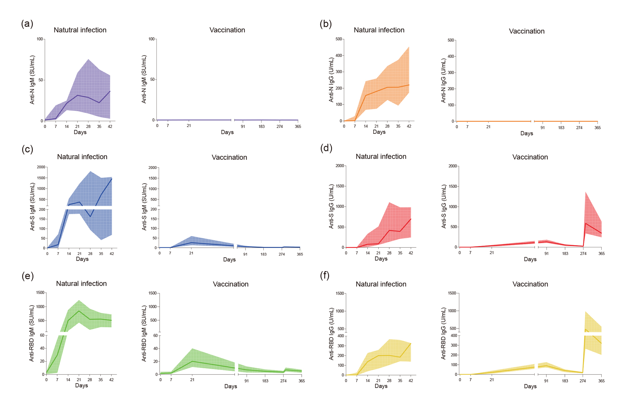
Figure 1 Longitudinal change in antibody titers of natural infection and vaccination.
In natural infection, a total of 197 sequential serum samples from 13 COVID-19 patients collected up to 47 days after symptom onset were analyzed. In vaccination, blood samples were collected from 25 SARS-CoV-2 naïve individuals who received a three-dose regimen of the BNT162b2 vaccine. Blood was collected at seven timepoints up to 365 days after the first-dose.
Anti-IgM titers against the N protein (a), the S protein (c), and the RBD (e); anti-IgG titers against the N protein (b);
the S protein (d), and the RBD (f) were determined by performing a CLEIA. Lines of the same color indicate median antibody titers with interquartile range. Manufacturer’s positive cut-off concentration sets at 20 SU/mL for IgM, 10 U/mL for IgG.
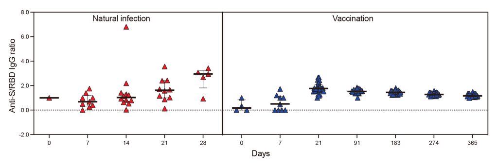
Figure 2 Comparison of anti-S/RBD IgG and IgM ratio in natural infection and vaccination.
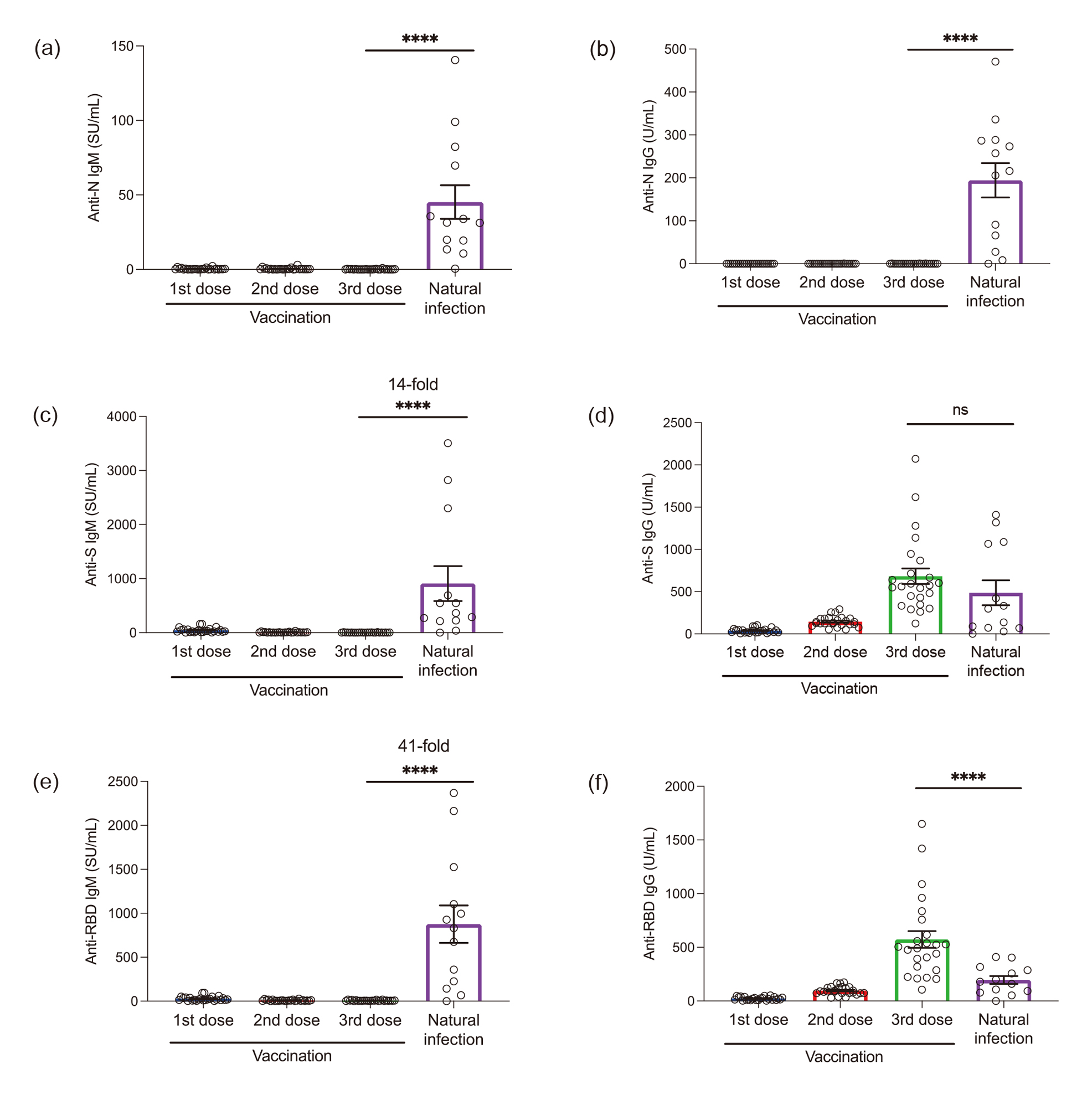
Figure 3 Comparative analysis of peak antibody titers to natural infection and the BNT162b2 mRNA vaccine.
Antibody responses to natural infection and vaccination were assessed by a CLEIA. Anti-IgM peak titers against the N protein (a), the S protein (c), and the RBD (e); anti-IgG peak titers against the N protein (b); the S protein (d), and the RBD (f) were determined. The P value (two-tailed) was calculated using the non-parametric Mann-Whitney U test. ns p>0.05, ****p<0.0001. Results represent individual values (dots) and means ± the SEM (bars) for anti-IgM and IgG antibodies recognizing N, S, and RBD SARS-CoV-2 antigens.
IV. Limitations of the study
We recognize some limitations in the present study. First, we included only 13 naturally infected and 25 vaccinated individuals; therefore, the sample size was small. In addition, the distribution of age groups in a population and the small sample size of women in the naturally infected group and men in the vaccinated group might have affected the analysis results. Second, information on adverse reactions was obtained using a self-reporting questionnaire. Thus, it was likely not accurate and was influenced by self-reporting bias. Third, we used the remaining serum after routine testing in patients who were naturally infected; thus, obtaining the same time points of sera among patients was challenging. Finally, the reactivity of the SARS-CoV-2 antibody reagents could vary based on the type of SARS-CoV-2 antibody produced by natural infection and vaccination. Despite these limitations, the present study provides evidence of the association between the target antigen and immunoglobulin isotypes and their characteristics.
Ethics declaration
This study was approved by the Nagoya University Hospital Ethics Committee (identification number:2020-0095, 2021-0040).
Acknowledgments
The authors thank all the participants of this study. We thank Sysmex for technical support and for providing the CLEIA tests. We especially thank our laboratory staff who collected the sample/data.
Author Contributions
Conceptualization, R.K., J.K., and A.S.; methodology and investigation, J.K. and R.K.; data analysis, J.K., R.K., A.S.; Sample collection, J.K., R.W., K.G., and Y.O.; writing—original draft preparation, J.K.; writing—review and editing, R.K., A.S., R.W., K.G., and Y.O.; supervision, T.M. All the authors have read and agreed to the published version of the manuscript.
Data availability statement
The corresponding author had full access to all the data in the study and all authors shared final responsibility for the decision to submit for publication. The datasets generated during the current study are available from the corresponding author on reasonable request.
Funding
This publication was supported by a grant from Sysmex Corporation.
Competing interests
All reagents used for the analysis were provided by Sysmex Corporation. The Sysmex Corporation had no control over the interpretation, writing, or publication of this work.
References
- World Health Organization: COVID-19 vaccine tracker and landscape.
https://www.who.int/publications/m/item/draft-landscape-of-covid-19-candidate-vaccines(Accessed August 5, 2023.) - Morinaga Y, Tani H, Terasaki Y, et al. Correlation of the commercial anti-SARS-CoV-2 receptor binding domain antibody test with the chemiluminescent reduction neutralizing test and possible detection of antibodies to emerging variants. Microbiol Spectr 2021; 9(3): e0056021.PubMed
- Gruell H, Vanshylla K, Weber T, et al. Antibody-mediated neutralization of SARS-CoV-2. Immunity 2022; 55(6): 925-44.PubMed
- Noval MG, Kaczmarek ME, Koide A, et al. Antibody isotype diversity against SARS-CoV-2 is associated with differential serum neutralization capacities. Sci Rep. 2021; 11(1): 5538.PubMed
- Yang, Y, Yang M, Peng Y, et al. Longitudinal analysis of antibody dynamics in COVID-19 convalescents reveals neutralizing responses up to 16 months after infection. Nat Microbiol 2022; 7(3): 423-33.PubMed
- Wang, Z, Muecksch F, Schaefer-Babajew D, et al. Naturally enhanced neutralizing breadth against SARS-CoV-2 one year after infection. Nature 2021; 595(7867): 426-31.PubMed
- Borremans B, Gamble A, Prager KC, et al. Quantifying antibody kinetics and RNA detection during early-phase SARS-CoV-2 infection by time since symptom onset. elife 2020; 9: e60122.
https://doi.org/10.7554/eLife.60122PubMed - Anand SP, Prévost J, Nayrac M, et al. Longitudinal analysis of humoral immunity against SARS-CoV-2 Spike in convalescent individuals up to 8 months post-symptom onset. Cell Rep Med 2021; 2(6): 100290.PubMed
- Noda K, Matsuda K, Yagishita S, et al. A novel highly quantitative and reproducible assay for the detection of anti-SARS-CoV-2 IgG and IgM antibodies. Sci Rep 2021; 11(1): 5198.PubMed
- Crawford KHD, Dingens AS, Eguia R, et al. Dynamics of neutralizing antibody titers in the months after severe acute respiratory syndrome coronavirus 2 infection. J Infect Dis 2021; 223(2):197-205.PubMed
- Bonifacio MA, Laterza R, Vinella A, et al. Correlation between In Vitro neutralization assay and serological tests for protective antibodies detection. Int J Mol Sci 2022; 23(17): 9566.PubMed
- Muecksch F, Wise H, Templeton K, et al. Longitudinal variation in SARS-CoV-2 antibody levels and emergence of viral variants: a serological analysis. Lancet Microbe 2022; 3(7): e493-502.PubMed
- Poon RW, Lu L, Fong CH, et al. Correlation between Commercial Anti-RBD IgG Titer and Neutralization Titer against SARS-CoV-2 Beta Variant. Diagnostics (Basel) 2021; 11(12): 2216.PubMed
- Hale M, Netland J, Chen Y, et al. IgM antibodies derived from memory B cells are potent cross-variant neutralizers of SARS-CoV-2. J Exp Med 2022; 219(9): e20220849.PubMed
- Pušnik J, König J, Mai K, et al. Persistent maintenance of intermediate memory B cells following SARS-CoV-2 infection and vaccination recall response. J Virol 2022; 96(15): e0076022.PubMed
- Upasani V, Rodenhuis-Zybert I, Cantaert T. Antibody-independent functions of B cells during viral infections. PLoS Pathog 2021; 17(7): e1009708.PubMed
- Fraussen J. IgM responses following SARS-CoV-2 vaccination: Insights into protective and pre-existing immunity. EBioMedicine 2022; 77: 103922.PubMed
- Aranburu A, Ceccarelli S, Giorda E, et al. TLR ligation triggers somatic hypermutation in transitional B cells inducing the generation of IgM memory B cells. J Immunol 2010; 185(12): 7293-301.PubMed
- Jeeva S, Kim KH, Shin CH, et al. An update on mRNA-based viral vaccines. Vaccines (Basel) 2021; 9(9): 965.PubMed
- Tas JMJ, et al. Antibodies from primary humoral responses modulate the recruitment of naive B cells during secondary responses. Immunity. 2022; 55(10): 1856-1871.e6. 10.1016/j.immuni.2022.07.020PubMed
- Ode H, et al. Molecular epidemiological features of SARS-CoV-2 in Japan, 2020-1. Virus Evol. 2022; 8(1): veac034. 10.1093/ve/veac034PubMed
- Yafei LIU, Nakazaki Y, Arase H. Infectivity-enhancing antibodies against SARS-CoV-2. Translational and Regulatory Sciences 2022; 4(1): 1-4.J-STAGE
- Liu Y, Soh WT, Kishikawa JI, et al. An infectivity-enhancing site on the SARS-CoV-2 spike protein targeted by antibodies. Cell 2021; 184(13): 3452-66.PubMed
- Wang H, Yan D, Li Y, et al. Clinical and antibody characteristics reveal diverse signatures of severe and non-severe SARS-CoV-2 patients. Infect Dis Poverty 2022; 11(1): 15.PubMed