Akane Hashizume*1, Harumi Saeki*1, Takeshi Nakazawa*2, Akira Saito*3, Kazunori Miyake*2 , Shigeki Tomita*1
Cite
Hashizume A, Saeki H, Nakazawa T, Saito A, Miyake A , Tomita S, Cutaneous abscess with disseminated Mycobacterium abscessus from the lung. Lab Med Int 2022; 1(1): 13-16. doi: 10.51041/lmi.1.1_13
Case Report
Lab Med Int 2022; 1(1): 13-16
Correspondence: Department of Pathology, Juntendo University Urayasu Hospital, Urayasu 279-0021, Japan.
E-mail: akane”@”juntendo-urayasu.jp
This paper was presented at the 66th Annual Meeting of Japanese Society of Laboratory Medicine.
Received June 29, 2020; accepted June 18, 2021
*1 Department of Pathology, Juntendo University Urayasu Hospital
*2 Department of Laboratory, Juntendo University Urayasu Hospital
*3 Department of Pathology, Tokyo Bay Urayasu Ichikawa Medical Center
ABSTRACT
A woman in her sixties presented to the dermatology department with erythematous nodules. Seventeen years earlier, she had been diagnosed with rheumatoid arthritis and treated with oral prednisolone. Three years later, she was also diagnosed with pulmonary non-tuberculous mycobacteriosis. Initially, we isolated Mycobacterium intracellulare from the bronchoscopy. However, 8 years later, the bacterial species changed to Mycobacterium abscessus. Skin biopsy was performed, and the culture was positive for Mycobacterium abscessus.
〔Lab Med Int 2022; 1(1): 13-16〕
Key Words
nontuberculous mycobacteriosis, Mycobacterium abscessus, cutaneous abscess
I. Introduction
The incidence of non-tuberculous mycobacteria (NTM) infections has been increasing in recent years. Among the NTM, Mycobacterium intracellulare (M. intracellulare) is a common cause of pulmonary infection. A majority of disseminated NTM infections occur in patients with a compromised immune status, such as infection with human immunodeficiency virus1). M. abscessus, a rapidly growing NTM, can cause skin infection. M. abscessus infection is resistant to numerous antibiotics and difficult to treat.
Herein, we describe a case of cutaneous abscess with disseminated M. abscessus from the lung.
II. Case report
A woman in her sixties presented to the dermatology department with erythematous nodules. The nodules were observed on bilateral legs 5 days after a follow-up computed tomography scan of the chest with contrast (Figure 1). Seventeen years earlier, she had been diagnosed with rheumatoid arthritis and treated with oral prednisolone (5 mg/day). Three years later, she was also diagnosed with pulmonary non-tuberculous mycobacteriosis. We initially isolated M. intracellulare from the bronchoscopy; however, 8 years later, the bacterial species changed to M. abscessus. Following the diagnosis of M. intracellulare infection, the patient was treated with clarithromycin, rifampicin, and ethambutol. A combination antibiotic therapy comprising oral clarithromycin, intravenous meropenem, and amikacin was initiated for the M. abscessus infection. However, pneumonia was refractory to the treatment. Three years prior to consultation, spinal fusion surgery was performed for an osteoporotic vertebral compression fracture. She was admitted to our hospital due to the presence of erythematous nodules and progressive pneumonia.
The laboratory data on admission were as follows: C-reactive protein 9.8 mg/dL; matrix metalloproteinase-3 187.4 ng/mL; and rheumatoid factor 244 IU/mL. Chest computed tomography revealed bilateral consolidation and reticular shadows (Figure 2).
The patient underwent a punch biopsy of her right leg; the culture was positive for rapidly growing mycobacteria (Figure 3A). Using DNA–DNA hybridization, the mycobacteria in clinical isolates were identified as M. abscessus. The minimum inhibitory concentrations were as follows: amikacin 16.0 µg/mL; kanamycin 16.0 µg/mL; clarithromycin >32.0 µg/mL; levofloxacin 32.0 µg/mL; streptomycin 32.0 µg/mL; rifampicin >32.0 µg/mL; ethambutol >128.0 µg/mL; ethionamide >16.0 µg/mL; and rifabutin 8.0 µg/mL. Histologically, extensive neutrophil infiltration with multinucleated Langhans giant cells throughout the dermis and the subcutaneous tissue was found (Figure 3B). However, there were no plump epithelioid cells detected. The results revealed non-caseating suppurative necrosis. Acid-fast bacilli were identified by Ziehl–Neelsen staining (Figure 3C).
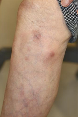
Figure 1 Appearance of the leg
III. Discussion
M. abscessus is a rapidly growing NTM and an environmental contaminant. It is regarded as chemotherapy-resistant NTM. M. abscessus can cause pulmonary and skin infections. In immunocompetent individuals, skin wounds are entry portals for M. abscessus. Disseminated infections, involving the lungs and skin, occur in immunocompromised patients. Guidelines established by the American Thoracic Society/Infectious Diseases Society of America (ATS/IDSA) recommend multidrug macrolide-based therapy based on susceptibility testing results1).
Occasionally, pleural M. abscessus infection develops during the treatment of non-abscessus mycobacterial disease2)-4). Culture examination should be considered in patients with NTM receiving long-term antibiotic therapy. A review of reported cutaneous M. abscessus cases showed various clinical characteristics5). Similar to our case, immunocompromised patients tend to have multiple lesions. M. abscessus has been associated with various foreign body infections6). In this case, metallic spinal implants may affect chronic infection with NTM. The ATS/IDSA recommended that surgery should generally be indicated for cases of M. abscessus infection with extensive disease, abscess formation, or where antibiotic therapy is difficult. Therefore, removal of foreign bodies is important and probably essential to recovery. In the present case, it was not possible to treat multiple lesions surgically or remove spinal implants, which may have increased the risk of chronic incurable infection.
Various types of bacteria and fungi cause granulomatous dermatitis. Because the treatment differs, the distinction of M. abscessus infection from other skin diseases is important. Combination antimicrobial therapy is recommended for disseminated M. abscessus infections. Differential diagnoses include cutaneous tuberculosis and swimming pool granuloma caused by M. marinum. M. marinum is an acid-fast bacillus, slightly larger than M. tuberculosis. M. abscessus is an elongated-shaped bacillus and, as the name implies, tends to from soft tissue abscesses. Sufficient clinical information is required for accurate pathological diagnosis.
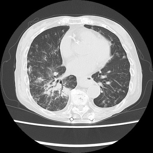
Figure 2 Chest computed tomography on admission showing bilateral consolidation and reticular shadows. Metallic spinal implants are visible.
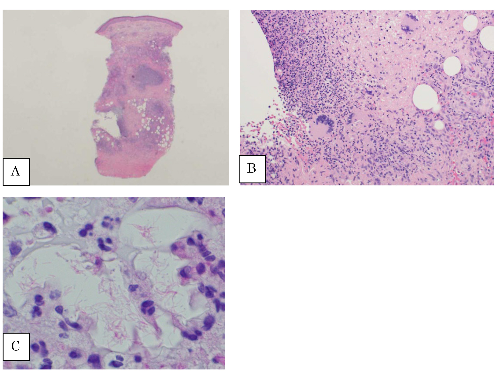
Figure 3
A) A low-power view of the skin (×1.25)
B) Abscess with multinucleated giant cells (×20)
C) Acid-fast bacilli shown by Ziehl–Neelsen staining (×100).
Conflict of interest
The authors declare that they have no conflicts of interest.
References
- Griffith DE, Aksamit T, Brown-Elliott BA, et al. An official ATS/IDSA statement: diagnosis, treatment, and prevention of nontuberculous mycobacterial diseases. Am J Respir Crit Care Med 2007; 175(4): 367-416. PubMed
- Tsunoda Y, Sekine A, Sumazaki Y, et al. Mycobacterium abscessus pulmonary disease: Important pathogen involved in microbial substitution during the treatment of non-abscessus mycobacterial disease. Kekkaku 2016; 91(4): 469-73. PubMed
- Griffith DE, Girard WM, Wallace RJ Jr. Clinical features of pulmonary disease caused by rapidly growing mycobacteria: An analysis of 154 patients. Am Rev Respir Dis 1993; 147(5): 1271-8. PubMed
- Nagano H, Amitani R, Okamoto N, et al. A clinical study of pulmonary Mycobacterium abscessus infection. Kansenshogaku Zasshi 2013; 87(6): 726-31. PubMed
- Koto M, Okazaki S, Fukano H, et al. A case of Mycobacterium abscessus subsp. massiliense skin disease. Rinsho derma. 2020; 62(7): 1053-8 (in Japanese).
- Tejura N, Bontempo G, Chew D. Disseminated Mycobacterium abscessus infection secondary to an infected vascular stent: Case report and review of the literature. Open Forum Infect Dis 2018; 5(9): ofy207. PubMe