
Norihito Morimoto, PhD*1,2, Yoshie Nishida, PhD*2, Masataka Korenaga, PhD*1,3
Morimoto N, Nishida Y, Korenaga M. Morphological and Genetic Analysis of Diploscapter coronatus: Insights into Identification Challenges and Species Relationships. Lab Med Int 2025; 4(2): 69-74. doi: 10.51041/lmi.4.2_69
Original
Lab Med Int 2025; 4(2): 69-74
Correspondence: Departments of Medical Laboratory Science, Kochi Gakuen University, 292-26 Asahitenjin-cho, Kochi 780-0955, Japan. E-mail: nmorimoto”@”kochi-gu.ac.jp.
Received November 21, 2024; accepted January 9, 2025
*1 Department of Medical Laboratory Science, Kochi Gakuen University
*2 Department of Clinical Laboratory, Kochi Medical School Hospital, Kochi University
*3 Department of Parasitology, Kochi University Medical School, Kochi University
ABSTRACT
Objective: This study aims to address the challenges of identifying Diploscapter coronatus by morphological and genetic analysis. The nematode was previously identified morphologically in fecal samples from a patient with Henoch-Schönlein purpura. We used genetic methods to complement and refine species identification.
Materials and Methods: Nematodes isolated from the patient’s feces were fixed in formalin, and morphological observations were made under a light microscope. PCR amplification of the SSU rRNA and Hsp90 genes was performed on DNA extracted from the nematodes during the same period. The resulting DNA was cloned, sequenced, and subjected to phylogenetic analysis using the neighbor-joining method.
Results: Morphological observations confirmed that the nematode shared key features with D. coronatus. The SSU rRNA analysis showed 99% similarity with 11 Diploscapter species, including D. coronatus. However, Hsp90 gene analysis placed the nematode in the D. lycostoma cluster, revealing inconsistencies between morphological and genetic data.
Conclusion: The present study highlights the complexity of species identification within the genus Diploscapter that results from the limited genetic information and overlapping morphological characters. The results underscore the need for more extensive genetic data and comprehensive phylogenetic analyses to resolve these identification challenges and better understand species relationships within the genus.
〔Lab Med Int 2025; 4(2): 69-74〕
Key Words
Diploscapter coronatus, Diploscapter spp., genetic analysis, small subunit ribosomal RNA (SSU rRNA), heat shock protein 90 (Hsp90)
I. Introduction
Diploscapter coronatus was first described by Cobb in 1893 as a free-living nematode typically found in soil surrounding decaying plants and roots1). Its detection in human samples is very rare, although its presence has been noted in past cases of anoxia and alkaline urine2), 3). In addition, recent reports have identified nematodes suspected of being this species in clinical samples such as urine and feces4), 5). Several species, including D. coronatus, have been documented within the genus Diploscapter. In 2002, we detected this nematode in fecal samples from a patient with Henoch-Schönlein purpura, identified by its morphological features6). Recently, both morphological and genetic methods have been used for species identification in nematode studies7). In this study, we report the analysis of a sample of D. coronatus, previously identified by morphological observations and now complemented by genetic identification methods.
II. Materials and Methods
1. Samples
Nematodes found in patient feces in 2002 were cultured in nematode growth medium (NGM), then fixed in 10% buffered formalin and stored at room temperature. Morphological observations were carried out on these formalin-fixed nematodes. During the same period, genomic DNA was extracted from D. coronatus nematodes derived from the patient and cultured in NGM using a QIAamp DNA Micro Kit (Qiagen, Hilden, Germany) and stored frozen at −80℃. This study was approved by the Ethics Committee of Kochi University Medical School, Japan (No.30-87).
2. Morphology
Morphological observations and measurements of the formalin-fixed nematodes were made using a light microscope and considered in conjunction with the observations we have previously reported6). Using De Man’s formula8), specific ratios and indices were calculated based on measurements of different parts of the nematode’s body to describe its morphological characteristics.
3. Polymerase Chain Reaction (PCR) and Cloning
The primers used for the small subunit ribosomal RNA (SSU rRNA) gene were obtained from Holterman et al9). (2006), and those for the 90 kDa heat shock protein (Hsp90) gene were obtained from Zeng Qi Zhao7) (2013), respectively. The 20 µL PCR reactions contained 10 µL KAPA2G Fast PCR Kit (Kapa Biosystems, USA), 1 µL (0.05 µM) of each forward and reverse primer, and 2 µL of DNA template. The thermal cycling program was as follows: denaturation at 95℃ for 3 min, followed by 30 cycles of denaturation at 94℃ for 30 s, annealing at 55℃ for 30 s, and extension at 72℃ for 45 s. A final extension was performed at 72℃ for 10 min. The amplified DNA was purified using a QIAGEN Gel Extraction Kit (QIAGEN, Valencia, CA, USA). The cDNA was ligated into a T-vector using the DynaExpress TA PCR Cloning Kit (BioDynamics Laboratory, Tokyo, Japan) and then transformed into competent Escherichia coli DH5α cells (FUJIFILM Wako, Tokyo, Japan) using the heat shock method. Finally, the recombinant plasmid was extracted from the transformed E. coli, using EZ-10 Spin Column Plasmid DNA Miniprep Kit (BIO BASIC, Ontario, Canada).
4. Sequencing and Phylogenetic Analysis
Purified plasmids were sequenced using the Big Dye™ Terminator Cycle Sequencing Ready Reaction Mix v3.1 Kit. Cycle sequencing products were purified using the FastGene Dye Terminator Removal Kit (NIPPON Genetics, Tokyo, Japan) and analyzed on an ABI PRISM 310 Genetic Analyzer (Applied Biosystems, Foster City, CA, USA). The genetic sequences obtained were subjected to similarity searches using BLAST10) from the National Center for Biotechnology Information (NCBI). DNA sequences were aligned using Clustal OMEGA11) and a phylogenetic tree was constructed using the Neighbor-Joining method with MEGA 1112). The root of the phylogenetic tree was constructed using the nucleotide sequence of Rhabditis myriophila (U81588) for SSU, and Acrobeloides amurensis (DQ340377) and Cephalobus cubaensis (DQ340378) for HSP90.
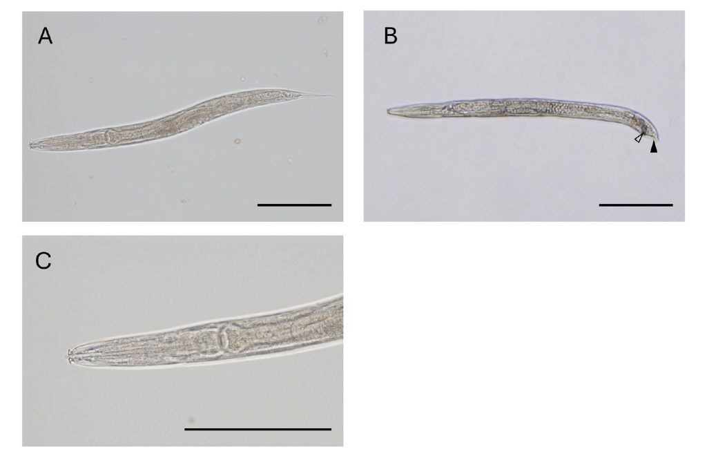
Figure1 Microscopic morphology of adult nematodes.
Whole body of both adult female( A) and male exhibiting the presence of spicules( △) and caudal alae( ▲) in the tail( B). Enlarged view of the head of an adult female( C). Scale bar indicating 100 μm.
III. Results
1. Morphological Observations
The microscopic images of the male and female nematodes in this study are shown in Figure 1. The length of the adult worms ranged from 344 to 522 µm, with a width of 22 to 39 µm (Table 1). They had two characteristic pairs of lips on the head, with the dorsal and ventral lips being hook-shaped and the second pair branching anteriorly, consistent with the characteristics of the genus Diploscapter (Figure 1B). To compare this nematode with the genus Diploscapter, coefficients calculated from the De Man measurements are presented in Table 1. Morphologically, both the size and the De Man ratios were similar to those of D. coronatus.
2. Genetic Findings
A homology search using BLAST showed that the SSU rRNA region of this nematode exhibited 99% similarity to 11 Diploscapter species. The similarity to the two included D. coronatus sequences was 99.3%. In the Hsp90 region, similarity ranged from 89.8% to 99.4% for D. lycostoma and from 86.8% to 89.7% for D. coronatus (Table 2). Phylogenetic analysis classified the Diploscapter species into clusters based on the four SSU regions, with the nematode in this study placed in a cluster that included D. coronatus (Figure 2). In contrast, using the Hsp90 data, the Diploscapter species were classified into clusters corresponding to D. coronatus, D. formicidae and D. lycostoma with the nematode in this study placed in the D. lycostoma cluster (Figure 3).
Table 1 Comparative measurements of adluts Diploscapter spp.*
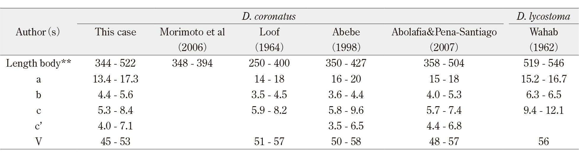
*The De Man ratios (a to c’+V) Favoured by plant and soil nematologists are as follows: a=body length/maximun width; b=body
length/length of esophagus; c=body length/tail length; V= Percentage of body length from the head to the vulva.
**Length body( μm).
Table 2 Homology search in the SSU rRNA region using BLAST
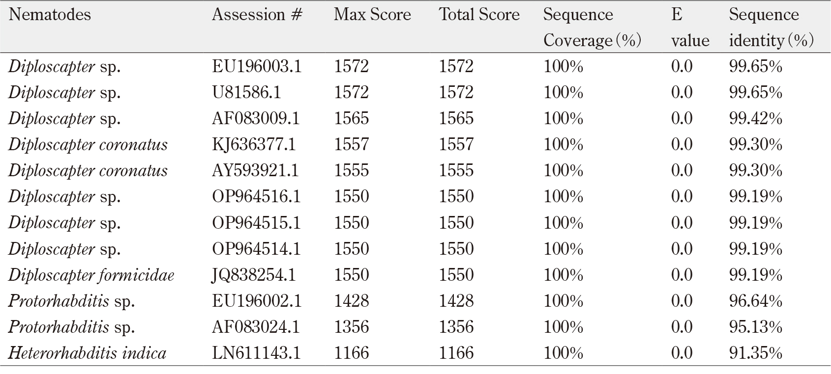
Homology search using BLAST was performed with the nucleotide sequences obtained from sequencing the plasmid
DNA, which contained the PCR product of the SSU region of this nematode.
IV. Discussion
Morphologically, it has been estimated that there are approximately 15 species within the genus Diploscapter13). However, to date, comprehensive genetic analyses and electron microscopic evaluations have not been carried out for most species. The nematode analyzed in this study matched the morphological characteristics of D. coronatus observed in previous studies14)-17). In addition, the b-ratio (body length/esophagus length) of D. coronatus differed from that of D. lycostoma in the De Man ratios referenced for nematodes17). From these findings, the nematode in this case demonstrated a b-ratio of 4.4-5.6, which morphologically identified it as D. coronatus.
The information available in NCBI on the genus Diploscapter is particularly limited, and only three species are fully documented: D. coronatus, D. lycostoma and the newly described D. formicidae. The nematode in this study showed high similarity to D. coronatus in the SSU rRNA gene, while in the Hsp90 region, it exhibited higher similarity to D. lycostoma. We currently lack sufficient genetic data on the SSU rRNA region in D. lycostoma. In addition, the genetic information available for the Hsp90 region lacks corresponding, detailed morphological descriptions. These factors likely contribute to the inconsistencies observed using genetic identification methods. In practice, when performing a BLAST search for the Hsp90 gene sequence of D. lycostoma (EF467042), the similarity to other D. lycostoma sequences ranges from 91.4% to 93.6%, and to D. coronatus sequences ranges from 85.8% to 85.9%. Identification by genetic methods is therefore challenging.
Interestingly, G. Markin et al18) reported that D. coronatus (Cobb 18931)) is synonymous with D. lycostoma (Volk 1950)19). The paucity of samples available for phylogenetic analysis may suggest that the resulting phylogenetic tree does not accurately reflect the true evolutionary relationships between the classified groups.
To date, while D. coronatus has been detected in human samples4), 5), there have been no reports of other species within the genus Diploscapter being detected in humans.
In addition, the detection of Diploscapter formicidae7) and Diploscapter lycostoma19), 20) in association with ants suggests a possible correlation between species and host specificity. In general, when a rare parasite is detected in humans, species identification is often performed based on the similarity of gene sequences from a particular genetic region, and genetic identification for various organisms has advanced in clinical examination. However, for the genus Diploscapter, the current genetic information available is insufficient, leading to uncertainties in species identification. In fact, Arai et al21) reported that although they attempted to genetically identify Diploscapter from human samples, a definitive identification of D. coronatus could not be achieved because of limited nucleotide sequence information.
Interestingly, although males are thought to be absent in the genus Diploscapter, we have reported the presence of males in this genus6). Furthermore, the presence of males in D. coronatus has also been reported by other researchers4), 22). If males could be found in other species of the genus Diploscapter, it might be possible to identify species based on characteristics such as male reproductive organs.
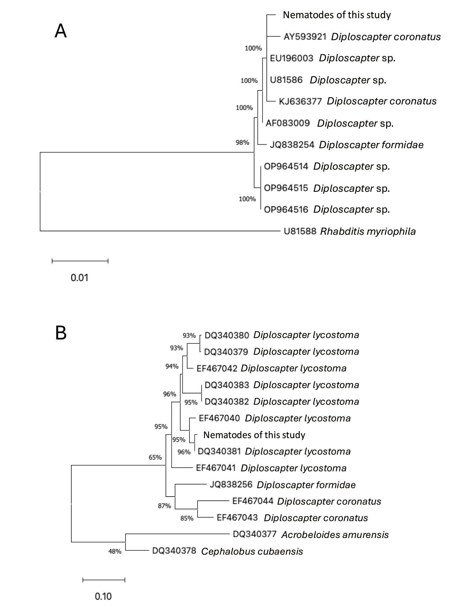
Figure 2 Neighbor-joining phylogenetic trees inferred from SSU rRNA gene( A) and HSP90 gene( B).
The phylogenetic trees include sequences from 9 species for SSU rRNA DNA and from 11 species for the
HSP90 gene within the genus Diploscapter, along with sequences from other related taxa. Bootstrap values are
shown at the nodes. The scale bar indicates the number of nucleotide substitutions per site. Sequences from
Rhabditis myriophila and other outgroup species were used to root the tree.
Table 3 Homology search in the HSP90 region using BLAST
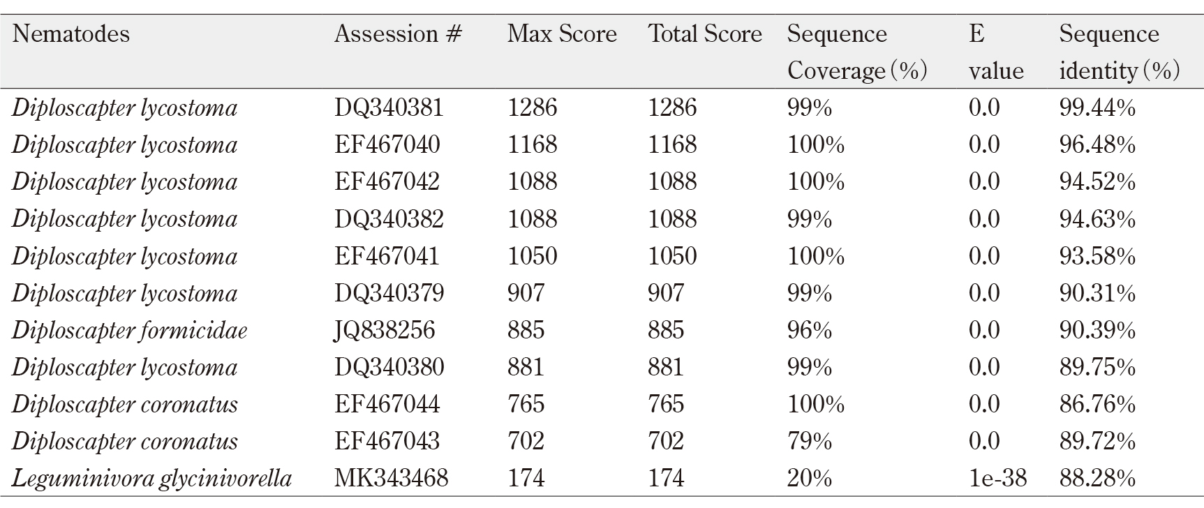
Homology search using BLAST was performed with the nucleotide sequences obtained from sequencing the plasmid
DNA, which contained the PCR product of the HSP90 region of this nematode.
V. Conclusion
The identification of species within the genus Diploscapter requires more extensive morphological and molecular data, with particular emphasis on comparing genetic information among different species to advance phylogenetic analyses. In addition, the ability to specifically detect species will likely provide important insights into host specificity.
VI. Acknowledgements including funding
We thank Bevin McGeever, PhD, from Edanz (https://jp.edanz.com/ac) for editing a draft of this manuscript.
Authorship contributions
All authors were involved in the preparation of the manuscript and reviewed the final manuscript.
Disclosure of Conflicts of Interest
We declare that we have no conflicts of interest.
References
- Cobb NA. 1893. Nematodes, mostly Australian and Fijian. Department of Agriculture, New South Wales. Miscellaneous Publication No. 13: 3-59.
- Chandler AC. Diploscapter coronata as a facultative parasite of Man, with a general review of vertebrate parasitism by rhabditoid worms. Parasitology. 1938; 30(1): 44-55.
- Yokogawa S. A human case of accidental parasitism of Diploscapter coronata (Cobb, 1893) Cobb, 1913. Zool Mag. 1936; 48: 507-11. (Japanese with English summary)
- Watthanakulpanich D, Anantaphruti MT, Maipanich W. Diploscapter coronata infection in Thailand: report of the first case. Southeast Asian J Trop Med Public Health. 2005; 36 (Suppl 4): 99-101.PubMed
- Athari A, Mahmoudi MR. Diploscapter coronata Infection in Iran: Report of the First Case and Review of Literature. Iranian J Parasitol. 2008; 3(3): 42-7. PubMed
- Morimoto N, Korenaga M, Yagyu K, et al. Morphological observations and the effects of artificial digestive fluids on the survival of Diploscapter coronata from a Japanese patient. J. Helminthol. 2006; 80(4): 341-48.PubMed
- Zhao ZQ, Davies KA, Brenton-Rule EC, et al. Diploscapter formicidae sp. n. (Rhabditida: Diploscapteridae), from the ant Prolasius advenus (Hymenoptera: Formicidae) in New Zealand. Nematology. 2013; 15(1): 109-23. doi:10.1163/156854112X649828.
- De Man JG. On the free-living nematoids and some other animals of brackish water and fresh water. Tijdschrift der Nederlandsche Dierkundige Vereeniging. 1880; 5: 1-103.
- Holterman M, van der Wurff A, van den Elsen S, et al. Phylum-wide analysis of SSU rDNA reveals deep phylogenetic relationships among nematodes and accelerated evolution toward crown clades. Mol Bio Evol. 2006; 23(9):1792-800. doi.org/10.1093/molbev/msl044.PubMed
- Altschul SF, Madden TL, Schäffer AA, et al. Gapped BLAST and PSI-BLAST: a new generation of protein database search programs. Nucleic Acids Res. 1997; 25(17): 3389-402. doi: 10.1093/nar/25.17.3389.PubMed
- Sievers F, Wilm A, Dineen D, et al. Fast, scalable generation of high−quality protein multiple sequence alignments using Clustal Omega. Mol Syst Biol. 2011; 7: 539. doi: 10.1038/msb. 2011.75PubMed
- Tamura K, Stecher G, Kumar S. MEGA11: Molecular Evolutionary Genetics Analysis Version 11. Mol Biol Evol. 2021; 38(7): 3022-27. doi: 10.1093/molbev/msab120.PubMed
- Sudhaus W. Phylogenetic systematisation and catalogue of paraphyletic “Rhabditidae” (Secernentea, Nematoda). J Nematode Morphol Syst. 2011; 14(2): 113-78.
- Loof PAA. Free-living and plant-parasitic nematodes from Venezuela. Nematologica. 1964; 10: 201-300.
- Abebe E, Karegar A, Nabil H, et al. A redescription and ultrastructural study of Diploscapter coronatus (Cobb, 1983) Cobb, 1913 from Ethiopia and Iran. Russ. J Nematol. 1998; 6(1): 17-22.
- Abolafia J, Peña-Santiago R. Nematodes of the order Rhabditida from Andalucía Oriental, Spain. The genera Protorhabditis (Osche, 1952) Dougherty, 1953 and Diploscapter Cobb, 1913, with description of P. spiculocrestata sp. n. and a species Protorhabditis key. J Nematol. 2007; 39(3): 263-74.
- Wahab A. Untersuchungen über Nematoden in den Drüsen des Kopfes der Ameisen (Formicidae). Zeitschrift für Morphologie und Ökologie der Tiere. 1962; 52(1): 33-92.
- Markin GP, McCoy CW. The Occurrence of a Nemotode, Diploscapter lycostoma, in the Pharyngeal Glands of the Argentine Ant, Iridomyrmex humilis. Ann Entomol Soc Am. 1968; 61(2): 505-9. doi.org/10.1093/aesa/61.2.505
- Völk J. Die Nematoden der Regenwürmer und aasbesuchenden Käfer. Zoologische Jahrbücher (Systematik) 1950; 79: 1-70. [Cited in: Tahseen Q, Siddiqi M.R, Rowe J. Study on species of Diploscapter Cobb (Nematoda: Rhabditida) from India including D. indicus sp. n. Int J Nematol. 2002; 12: 183-88.
- Lynn C, Andrea S, Eric E. Low temperature SEM and molecular phylogenetics of three Diploscapter populations. J Nematol 2006; 38(2): 36.
- Arai M, Nishimura K, Miyauchi H, et al. A large number of nematodes found in the urinary sediment of a 79 years-old man. Clin Parasitol. 2015; 26(1): 39-42. (in Japanese)
- Shah AA, Vaid S. Description of Sclerorhabditis miniata n. sp. and First Description of Male of Diploscapter coronatus Cobb, 1913 (Nematoda: Rhabditidae). J Nematol. 2015; 47(2): 153-8.PubMed