Kayo Yamada*1†, Alafate Ayibieke, PhD*1†, Akari Ikeda*1, Kageto Yamada, PhD*2, Shinji Ogihara, PhD*3, Shuji Tohda, MD, PhD*4, †Ryoichi Saito, PhD*1
Cite
Yamada K, Ayibieke A, Kurokawa K, Ikeda A, Yamada K, Ogihara S, Tohda S, Saito R. Molecular characterization of Staphylococcus epidermidis bloodstream isolates from two hospitals in Tokyo. Lab Med Int 2024; 3(2): 33-41. doi: 10.51041/lmi.3.2_33
Original
Lab Med Int 2024; 3(2): 33-41
† Correspondence: Department of Molecular Microbiology, Graduate School of Medical and Dental Sciences, Tokyo Medical and Dental University(TMDU), 1-5-45, Yushima, Bunkyo-ku, Tokyo 113-8510, Japan.
E-mail: r-saito.mi”@”tmd.ac.jp
Received February 6, 2023; accepted April 26, 2024
*1 Department of Molecular Microbiology, Tokyo Medical and Dental University Graduate School of Medical and Dental Sciences, Tokyo, Japan
*2 Department of Microbiology and Infectious Disease, Toho University School of Medicine, Tokyo, Japan
*3 Department of Clinical Laboratory, Toho University Medical center Omori Hospital, Tokyo, Japan
*4 Department of Clinical Laboratory, Tokyo Medical and Dental University Hospital, Tokyo, Japan
† These two authors contributed equally to this work.
ABSTRACT
Dissemination of multidrug-resistant Staphylococcus epidermidis, especially methicillin-resistant S. epidermidis (MRSE), with enhanced pathogenicity is a serious global public health concern. We characterized the antimicrobial/biocide susceptibility and virulence of 105 S. epidermidis bloodstream isolates from two hospitals in Tokyo to expand on the limited information available in Japan.
The phenotypic and genetic features of antimicrobial resistance, biocide tolerance, SCCmec type, and biofilm development or adhesion were analyzed.
In total, 76 (72.4%) isolates were identified as MRSE, which showed higher resistance rates to most antimicrobial classes, except for vancomycin, than methicillin-susceptible S. epidermidis (MSSE). MRSE was classified into 16 sequence types (STs), including the most prevalent ST2, which is a global high-risk S. epidermidis clone. In addition, our MRSE isolates possessed higher rates of qacA/B than the MSSE isolates, resulting in a higher tolerance to the three low-level antiseptics compared to MSSE isolates. Among these, ST6 and ST2 isolates showed higher qacA/B positivity rates. Furthermore, SCCmec type IV was predominant in the MRSE isolates. MRSE isolates possessed aap and IS256 more frequently than MSSE isolates. Moreover, sesI and icaA were found in the ST2 isolates. The proportion of biofilm producers in MRSE tended to be higher than that in MSSE, and strong biofilm producers were concentrated in ST2 isolates among the four predominant STs.
Collectively, our findings provide the first evidence that highly virulent, multidrug-resistant S. epidermidis isolates, including the global ST2 MRSE lineage, may have already spread and persisted for a long time in healthcare facilities in Japan.
〔Lab Med Int 2024; 3(2): 33-41〕
Key Words
Staphylococcus epidermidis, methicillin resistance, mecA, biocide tolerance, biofilm, sequence type
I. Introduction
Staphylococcus epidermidis is the most common species of coagulase-negative staphylococci (CoNS) and a normal human commensal bacterium in the skin and mucous membranes. Although it is an important opportunistic pathogen, it is also a leading cause of healthcare-associated infections (HAIs) involving medical devices such as catheters and prosthetic joints1)2). In Japan, S. epidermidis is the most frequently identified pathogen associated with catheter-associated bloodstream infections3).
S. epidermidis can acquire determinants conferring antimicrobial resistance and biocide tolerance, which can be horizontally transferred to other Staphylococcus species1)4). The recent emergence and dissemination of multidrug-resistant S. epidermidis, especially methicillin-resistant S. epidermidis(MRSE) with staphylococcal cassette chromosome mec(SCCmec), has become a global concern because of the difficulty in treating infections, prolonged hospitalization, and increased healthcare costs5). In addition, S. epidermidis has enhanced pathogenicity due to acquired virulence factors, such as biofilm development and adherence, and has been isolated globally2)6). Furthermore, among several global hospital-adapted HAI-associated S. epidermidis lineages, sequence type(ST) 2 of clonal complex 2(CC2) has been one of the most prevalent MRSEs in recent decades 2)5)7).
Although these factors may have contributed to the MRSE lineage becoming a globally successful clone in healthcare facilities, this phenomenon is not yet fully understood. Despite the frequent isolation of HAIs globally, knowledge regarding the phenotypic and genomic characteristics, such as antimicrobial resistance or virulence, of S. epidermidis isolates from Japan is still limited. Therefore, in this study, we aimed to clarify the profiles of antimicrobial and biocidal susceptibilities, biofilm formation, and genetic features, including the SCCmec type, of S. epidermidis bloodstream isolates from Japan.
II. Methods
A. Bacterial samples and growth conditions
A total of 105 non-duplicate S. epidermidis isolates recovered from at least two sets of positive blood cultures per patient, collected between 2007 and 2015 (n = 43) at the Tokyo Metropolitan Toshima Hospital and between 2014 and 2015 (n = 62) at the Tokyo Medical and Dental University Hospital, were used in this study. Microbial identification was confirmed using a MALDI Biotyper (Bruker Daltonics, Karlsruhe, Germany). The S. epidermidis strains were grown at 37℃ under aerobic conditions on tryptone soya agar plates and tryptone soya broth (Oxoid, Hampshire, United Kingdom).
B. Antimicrobial susceptibility testing and minimal inhibitory concentrations of biocides
Minimal inhibitory concentrations (MICs) of 12 antimicrobials (penicillin, oxacillin, ampicillin, cefazolin, imipenem, gentamicin, erythromycin, clindamycin, minocycline, levofloxacin, vancomycin, and sulfamethoxazole/trimethoprim), were determined by microdilution using commercial plates (Eiken Chemical, Tokyo, Japan). The detailed MIC of oxacillin in all ST2 isolates was also measured using Etest (bioMérieux Marcy-l’Étoile, France). All the results were interpreted according to the CLSI M100- ED30 guidelines.
Similarly, the MICs of chlorhexidine, benzalkonium chloride, and olanexidine gluconate were determined using the broth doubling microdilution method according to the CLSI M100-ED30 guidelines8).
C. Antimicrobial resistance, biocide tolerance, and biofilm development or adhesion genes screening
DNA was extracted using the Cica GeneusTM DNA Extraction Reagent (Kanto Chemical, Tokyo, Japan). mecA and seven biofilm development- or adhesion-associated genes (sesI, icaA, bhp, aap, atlE, arcA, and IS256) identified in Staphylococcus species were screened using PCR as previously described9)-14). PCR screening was also conducted for two major multidrug efflux pump-encoding genes for biocide tolerance: qacA/B and smr15).
D. Multi-locus sequence typing
Multilocus sequence typing (MLST) was performed using seven loci (arcC, aroE, gtr, mutS, pyrR, tpi, and yqiL), as previously described16), with some modifications. Briefly, aroE was amplified with the primers aroE-F2(5’-TCAGCACCTTGATGAACGAA-3) and aroE-R2(5’-GAACGTATTATTCCGTACCTAGATG-3). PCR was performed using the 2×EmeraldAmp MAX PCR Master Mix(Takara Bio, Shiga, Japan), and the products were purified and sequenced. STs were defined using the PubMLST S. epidermidis genome database (https://pubmlst.org/organisms/staphylococcus-epidermidis). The minimum spanning tree for genetic relatedness of the MLST data was visualized with goeBURST Full MST using PHYLOVIZ 2.017).
E. PCR-based SCCmec typing
PCR-based SCCmec typing of 76 MRSE strains was performed according to previously described methods 18)19), and the SCCmec type was interpreted based on the guidelines of the International Working Group on the Classification of Staphylococcal Cassette Chromosome Elements20). When the combined results for mec class and ccr type were not reported before, or when no mec or ccr was detected, they were classified as non-typeable.
F. Biofilm formation quantification
Biofilm formation assays were conducted in 96-well polystyrene plates (AS ONE Corporation, Osaka, Japan), as previously described21)22), with some modifications. Briefly, 3-h pre-cultured S. epidermidis isolates were inoculated into 2 ml fresh 1% glucose containing TSB (OD550 = 0.01–0.04), and then 200 µL of these cultures were transferred into each well, followed by overnight incubation at 37℃. Following incubation, all liquids were removed and the wells were washed three times with distilled water. The plates were stained with 1% (w/v) crystal violet solution, washed three times, and dried for 10–15 minutes. Finally, 200 µL 99.5% ethanol was added to each well, and the stained biofilms were measured at 570 nm using a MULTISKAN FC (Thermo Fisher Scientific, Waltham, MA, USA). Each isolate was tested at least six times; the mean results are presented.
Biofilm formation was assessed as described previously23). Briefly, the isolates were categorized as strong, moderate, weak, or non-biofilm producers according to the following equations:
OD < ODC (average OD of negative control) = non-biofilm producer
ODC < OD ≤ (2 × ODC) = weak biofilm producer
(2ODC) < OD ≤ (4 × ODC) = moderate biofilm producer
(4 × ODC) < OD = strong biofilm producer.
S. epidermidis ATCC 12228, which lacks biofilm formation, was used as a negative control.
G. Statistical analysis
Categorical variables were evaluated using the chi-square test. Statistical differences were determined using the Mann-Whitney U test. p < 0.05 was set as the threshold of significance.
III. Results
A. Antimicrobial susceptibility profile and its relationship with mecA prevalence
Among 105 S. epidermidis isolates, 76 (72.4%) were identified as MRSE, and 29 (27.6%) were identified as methicillin-susceptible S. epidermidis (MSSE)(Table 1). All MRSE isolates harbored mecA, whereas the MSSE isolates did not. The resistance rates to most other antimicrobial classes were significantly higher in MRSE than in MSSE. No vancomycin resistance was detected in any isolate.
The 105 isolates were classified into 33 STs, including six newly assigned STs (ST870, ST873, ST874, ST875, ST876, and ST891)(Figure 1). Twenty-nine MSSE isolates were more diverse and were classified into 21 STs while the 76 MRSE isolates were classified into 16 STs. ST5 (n = 21, 20%) was the most prevalent of 33 STs, followed by ST2 (n = 19, 18%), ST59 (n = 15, 14%) and ST6 (n = 11, 10%). Among these four STs, MRSE isolates accounted for the largest proportion (100% for ST2, 86% for ST5, 91% for ST59, and 73% for ST6).
B. Biocide MIC profiles and their association with STs and biocide tolerance genes
We investigated the relationship between the biocide MICs and STs. The geometric mean MICs of chlorhexidine, benzalkonium chloride, and olanexidine gluconate in the MRSE isolates were significantly higher than those in the MSSE isolates (Table 2). The prevalence of qacA/B was higher among the MRSE isolates (n = 39, 51%) than among the MSSE isolates (n = 3, 10%), whereas the prevalence of smr was similar between the MRSE and MSSE isolates (Table 3). ST6 and ST2 showed higher qacA/B positive rates of 100% and 53%, respectively, whereas the lowest frequencies of smr were observed in these two STs (9% and 11%, respectively) (Figure 2A).
The correlation between biocidal MICs and tolerance genes revealed that qacA/B-positive isolates showed significantly higher MICs for the three biocides than qacA/B-negative isolates (Figure 2B). However, smr-positive isolates exhibited significantly higher MIC for benzalkonium chloride.
C. SCCmec analysis and its association with STs
Our 76 MRSE isolates were classified into 15 different types of SCCmec including non-typeable types. Among the SCCmec elements detected and classified, SCCmec Type Ⅳ(2B) (n = 29, 38%) was predominant, followed by Type Ⅳ (2B&5) (n = 11, 14%) (Table 4). However, 10 out of 15 different SCCmec types were classified as non-typeable SCCmec due to an unclassified combination of mec class and ccr type, and comprised 31 MRSE isolates (41%). Moreover, in terms of SCCmec types among the four major STs, the SCCmec types of ST6 isolates tended to be less diverse than those of the ST2, ST5, and ST59 isolates (Table 4).
D. Prevalence of biofilm development or adherence-associated genes
Next, we evaluated the frequency of virulence-associated genes, as S. epidermidis possesses several factors that are responsible for adhesion to host cells and biofilm formation. Among the MRSE strains, the frequency of aap, which is related to biofilm development through accumulation on polymer surfaces, and IS256, which is involved in biofilm production through the inactivation of ica operon-mediated phase variation by its insertion into S. epidermidis, was significantly higher than that of MSSE (Table 3). However, there was no significant difference in the prevalence of the other five genes between MRSE and MSSE.
sesI, which functions in cell-to-cell adhesion through the synthesis of a polysaccharide intercellular adhesin, and icaA, which is composed of the ica operon, were found in ST2 isolates (58% and 100%, respectively) (Figure 3). All ST2 strains were negative for bhp, a cell-wall anchored protein, whereas higher frequencies were found in ST5 and ST59 (52% and 67%, respectively). There were higher frequencies of aap among the three major strains, ST5, ST2, and ST59, whereas the prevalence of arcA, which encodes an arginine catabolic mobile element responsible for facilitating staphylococcal colonization of the skin and mucous membranes, was higher in the minor STs than in the three major STs. atlE, which mediates attachment to the polystyrene surface, was present in all isolates. Interestingly, ST6 was 100% positivity for IS256 and 100% negativity for icaA, aap, bhp, and arcA.
E. Biofilm development ability
We further characterized the biofilm phenotypes of the 105 S. epidermidis isolates. Although the prevalence of strong producers in MRSE strains tended to be higher than that in MSSE, the total ratio of isolates with weak to strong biofilm formation was not significantly different between MRSE and MSSE (p > 0.05, Figure 4A).
Strong producers were concentrated in ST2 isolates among the four predominant ST groups but were also found in other STs (Figure 4B). Interestingly, all ST6 and many ST59 isolates were categorized as non-biofilm producers, indicating that there were different distributions of biofilm phenotypes among each ST.
Table 1 Antibiotic susceptibility profiles of 105 S. epidermidis isolates
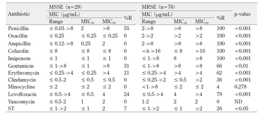
ST, sulfamethoxazole-trimethoprim. R, resistant. ND, not determined.
%R categorical variables were evaluated by the Chi-square test.
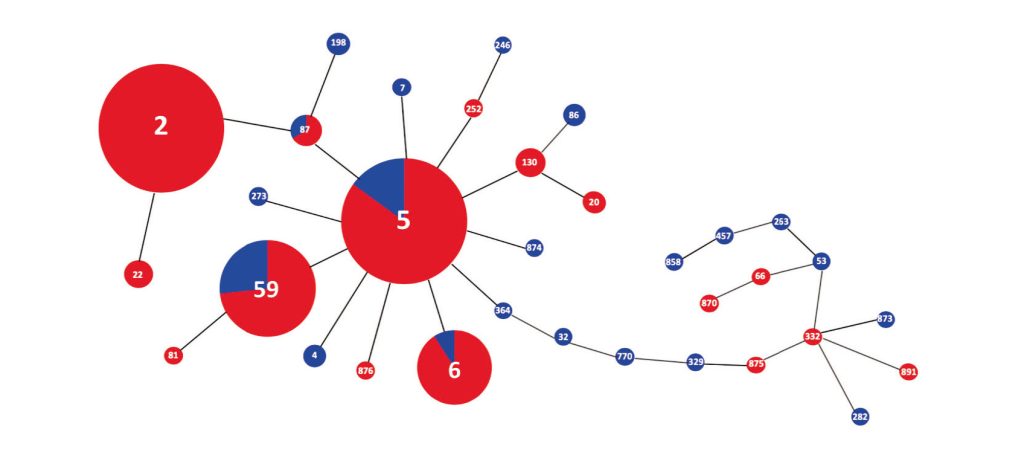
Figure 1 Minimum spanning tree based on the allelic profiles of 33 STs identified in 105 S. epidermidis isolates. Node sizes are proportional to the number of isolates for each ST and the numbers on connecting lines indicate the number of locus variants determined by pair-wise comparison. Red and blue areas represent MRSE and MSSE isolates, respectively.
Table 2 Biocide susceptibility profiles of 105 S. epidermidis isolates
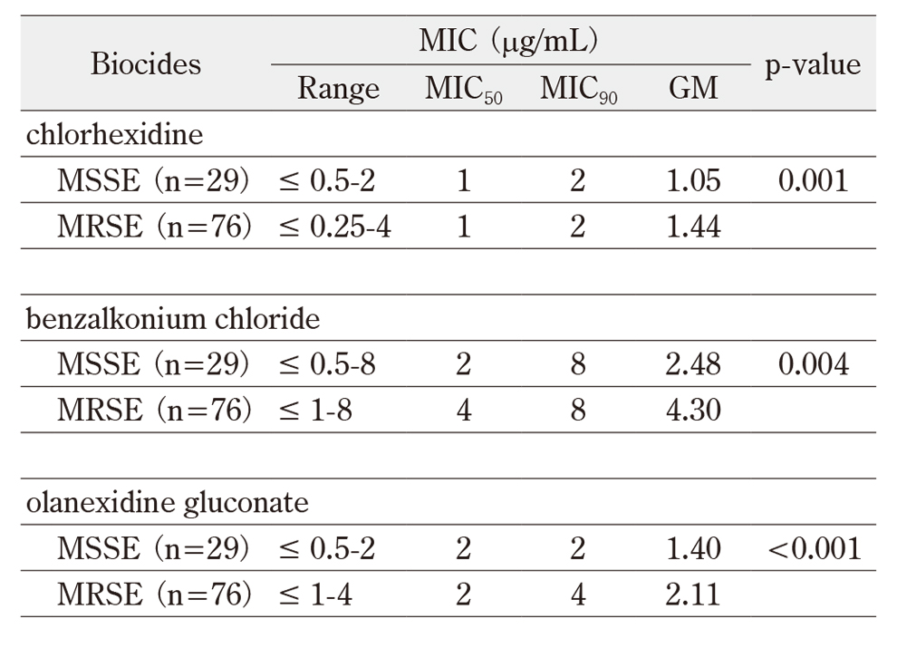
GM, geometric mean of MICs. GMs were compared using the Mann-Whitney U-test.
Table 3 Prevalence of biocide resistance and biofilm development/adhesion-associated genes of 105 S. epidermidis isolates
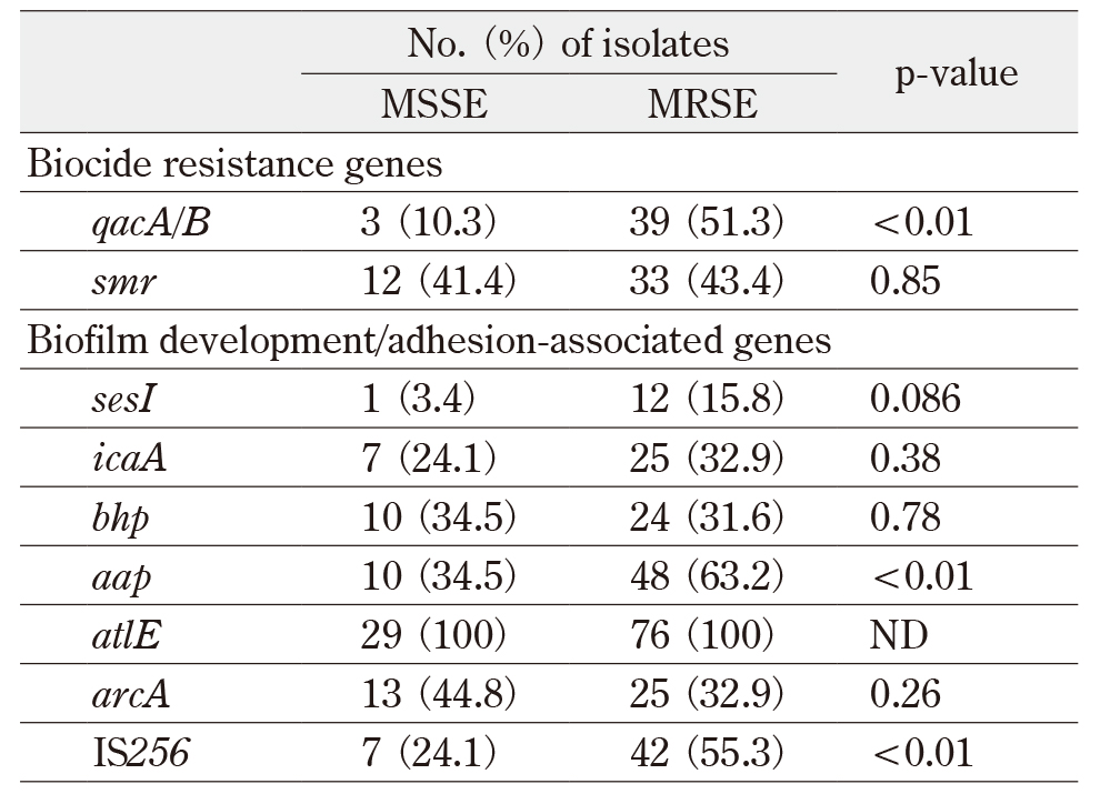
ND, not determined. Categorical variables were evaluated by the Chi-square test.
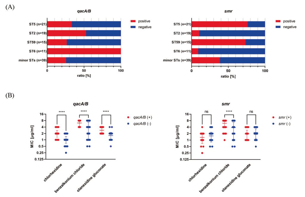
Figure 2 Prevalence of multidrug efflux pump-encoding genes and their association with biocide tolerance in 105 S. epidermidis isolates. (A) Prevalence of qacA/B and smr among each ST, and (B) the relationship between the presence of qacA/B and smr and three biocide MICs. Geometric means of biocide MICs were compared using the Mann-Whitney U-test. ****p < 0.0001.
Table 4 SCCmec profiles and its association with STs
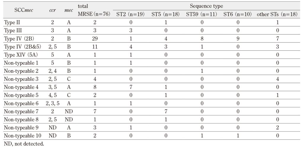
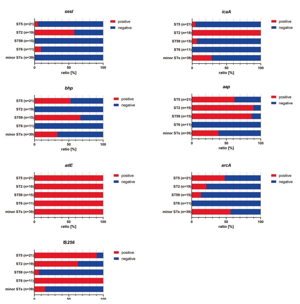
Figure 3 Prevalence of major biofilm development- or adhesion-associated genes among each ST. sesI, icaA, bhp, aap, atlE, arcA, and IS256 identified in Staphylococcus species were screened in 105 S. epidermidis isolates, as described in section C of Methods.
IV. Discussion
S. epidermidis clinical isolates tend to be multidrug-resistant, with oxacillin resistant-MRSE ranging from 70–90% prevalence, including data from nationwide surveillance in Japan1)3)24). Our results agree with this, highlighting that MSSE remains highly susceptible to several antimicrobials and that vancomycin is still recommended for treating MRSE infections. Our findings support the need to monitor antimicrobial resistance to effectively treat S. epidermidis infections.
S. epidermidis, especially MRSE, is a major reservoir of antimicrobial and biocidal resistance determinants4). In this study, MRSE isolates possessed higher rates of qacA/B than MSSE isolates and were associated with a higher tolerance to three low-level antiseptics. These results, coupled with those of previous studies on Staphylococcus species2)25), demonstrate that antimicrobial and antiseptic co-resistant/tolerant isolates may be spreading in Japanese hospitals. However, as QacA/B and Smr cannot functionally exhaust many intermediate-level antiseptics/disinfectants that are often used in healthcare facilities, such as ethanol26), from inside bacterial cells, appropriate antiseptics/disinfectants may be efficient in eliminating antiseptic/disinfectant-tolerant isolates.
Methicillin-resistant CoNS, including S. epidermidis, possesses multiple SCCmec elements, resulting in diverse structural variants19)27). Consistent with previous reports19)27), our results demonstrated that diverse SCCmec elements, including SCCmec type IV, were predominant, suggesting that CoNS in Japan may constitute a reservoir of SCCmec with methicillin resistance. Moreover, a previous study reported that SCCmec islands could evolve through site-specific recombination at this locus during adaptation to the human host28). Therefore, we may need to characterize SCCmec islands at the whole-genome level to clarify the global diversification of S. epidermidis used in this study.
We demonstrated that MRSE isolates frequently possessed the biofilm-forming or adherence-associated genes aap and IS256; these higher rates did not result in significant differences in phenotypic biofilm formation between MRSE and MSSE. This may be because major biofilm development- or adhesion-associated genes were not fully upregulated in the planktonic cells used in this study or because other determinants regulating the biofilm production process were present in our isolates. However, coupled with the results of antimicrobial/antiseptic co-resistance/tolerance, our findings suggest that highly pathogenic multidrug-resistant S. epidermidis isolates have already been disseminated in Japan. In addition, a previous study from China demonstrated that ST2 isolates were exclusively ica- and IS256-positive and biofilm-forming6). Our results showed a similar tendency, indicating for the first time that ST2 MRSE isolates might have higher pathogenicity among the four major STs in our isolates. Extracellular biofilms inhibit the action of most antimicrobials, thereby impairing the treatment of Staphylococcus medical-device infections. This indicates that the consistent application of infection prevention measures at an appropriate point, such as the periprocedural period, is crucial, in concordance with previous studies2)13). Moreover, previous studies have demonstrated that exopolysaccharides, such as polysaccharide intercellular adhesins, protect S. epidermidis from host defense mechanisms such as neutrophil killing1)29). Therefore, further investigation is needed to elucidate the relationship between STs, prevalence of virulence factors, and pathogenicity.
Among global high-risk S. epidermidis clones, ST2 belongs to the CC2 strain, which is the most widespread healthcare-associated strain2)5)6). However, there are limited reports focusing on the genetic features of ST2 MRSE, none of which are from Japan. The results of our MLST analysis provide novel insights that multidrug-resistant S. epidermidis CC2 isolates, including ST2 and ST5, may already be predominant and have adapted to healthcare facility environments in Japan. Further detailed genetic analysis using whole-genome sequencing may contribute to the understanding of genetic diversity and features that remain unidentified in antimicrobial resistance, virulence, and pathogenicity in CC2 MRSE healthcare-associated strains in Japan.
The current study had some limitations. For example, the number of clinical isolates collected from the two hospitals was small, and they were not compared with the isolates from healthy participants, and detailed information such as the isolated numbers and wards of this organism involved in horizontal transmission in each hospital was not investigated.
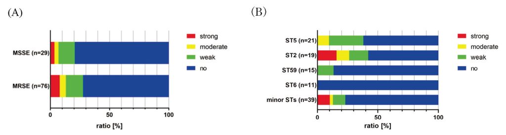
Figure 4 Biofilm development ability of 105 S. epidermidis isolates. (A) Prevalence of each biofilm phenotype in MSSE and MRSE isolates, and (B) relationship between the biofilm phenotype and STs. The phenotype was categorized, as described in section D of Methods.
V. Conclusion
Highly virulent, multidrug-resistant S. epidermidis isolates have emerged in Japan, complicating infection prevention and treatment. Our findings provide novel insights and reveal that the global ST2 MRSE clone and its close genetic lineages may have already been disseminated and have persisted for a long time in Japanese healthcare facilities. Therefore, robust surveillance systems and infection control and prevention measures based on continuous monitoring of their lineages are crucial for their elimination.
Acknowledgement
This study was supported by the Japanese Society of Laboratory Medicine Fund for the Promotion of Scientific Research. We would like to thank Editage (www.editage.jp) for English language editing.
Disclosures
We declare that we have no conflicts of interest. All authors read and approved the final manuscript.
References
- Otto M. Staphylococcus epidermidis–the ‘accidental’ pathogen. Nat Rev Microbiol 2009; 7(8): 555-67.PubMed
- Månsson E, Bech Johannesen T, Nilsdotter-Augustinsson Å, et al. Comparative genomics of Staphylococcus epidermidis from prosthetic-joint infections and nares highlights genetic traits associated with antimicrobial resistance, not virulence. Microb Genom 2021; 7(2).PubMed
- Japan Nosocomial Infections Surveillance Ministry of Health, Labour and Welfare, https://janis.mhlw.go.jp/english/report/index.html, (assessed Dec 20, 2022).
- Otto M. Coagulase-negative staphylococci as reservoirs of genes facilitating MRSA infection: Staphylococcal commensal species such as Staphylococcus epidermidis are being recognized as important sources of genes promoting MRSA colonization and virulence. Bioessays 2013; 35(1): 4-11.PubMed
- Lee JYH, Monk IR, Gonçalves da Silva A, et al. Global spread of three multidrug-resistant lineages of Staphylococcus epidermidis. Nat Microbiol 2018; 3(10): 1175-85.PubMed
- Li M, Wang X, Gao Q, et al. Molecular characterization of Staphylococcus epidermidis strains isolated from a teaching hospital in Shanghai, China. J Med Microbiol 2009; 58(Pt 4): 456-61.PubMed
- Miragaia M, Thomas JC, Couto I, et al. Inferring a population structure for Staphylococcus epidermidis from multilocus sequence typing data. J Bacteriol 2007; 189(6): 2540-52.PubMed
- Clinical and Laboratory Standards Institute. Performance Standards for Antimicrobial Susceptibility Testing: 30th Edition M100-ED30. CLSI, Wayne, PA, USA, 2020.
- Pérez-Roth E, Claverie-Martín F, Villar J, et al. Multiplex PCR for simultaneous identification of Staphylococcus aureus and detection of methicillin and mupirocin resistance. J Clin Microbiol 2001; 39(11): 4037-41.PubMed
- Bowden MG, Chen W, Singvall J, et al. Identification and preliminary characterization of cell-wall-anchored proteins of Staphylococcus epidermidis. Microbiology(Reading). 2005; 151(Pt 5): 1453-64.PubMed
- Najar Peerayeh S, Jazayeri Moghadas A, Behmanesh M. Prevalence of Virulence-Related Determinants in Clinical Isolates of Staphylococcus epidermidis. Jundishapur J Microbiol 2016; 9(8): e30593.PubMed
- Møretrø T, Hermansen L, Holck AL, et al. Biofilm formation and the presence of the intercellular adhesion locus ica among staphylococci from food and food processing environments. Appl Environ Microbiol 2003; 69(9): 5648-55.PubMed
- Du X, Zhu Y, Song Y, et al. Molecular analysis of Staphylococcus epidermidis strains isolated from community and hospital environments in China. PLoS One 2013; 8(5): e62742.PubMed
- Gu J, Li H, Li M, et al. Bacterial insertion sequence IS256 as a potential molecular marker to discriminate invasive strains from commensal strains of Staphylococcus epidermidis. J Hosp Infect 2005; 61(4): 342-8.PubMed
- Ho CM, Li CY, Ho MW, et al. High rate of qacA– and qacB-positive methicillin-resistant Staphylococcus aureus isolates from chlorhexidine-impregnated catheter-related bloodstream infections. Antimicrob Agents Chemother 2012; 56(11): 5693-7.PubMed
- Thomas JC, Vargas MR, Miragaia M, et al. Improved multilocus sequence typing scheme for Staphylococcus epidermidis. J Clin Microbiol 2007; 45(2): 616-9.PubMed
- Nascimento M, Sousa A, Ramirez M, et al. PHYLOViZ 2.0: providing scalable data integration and visualization for multiple phylogenetic inference methods. Bioinformatics 2017; 33(1): 128-9.PubMed
- Kondo Y, Ito T, Ma XX, et al. Combination of multiplex PCRs for staphylococcal cassette chromosome mec type assignment: rapid identification system for mec, ccr, and major differences in junkyard regions. Antimicrob Agents Chemother 2007; 51(1): 264-74.PubMed
- Ruppé E, Barbier F, Mesli Y, et al. Diversity of staphylococcal cassette chromosome mec structures in methicillin-resistant Staphylococcus epidermidis and Staphylococcus haemolyticus strains among outpatients from four countries. Antimicrob Agents Chemother 2009; 53(2): 442-9.PubMed
- Classification of staphylococcal cassette chromosome mec (SCCmec): guidelines for reporting novel SCCmec elements. Antimicrob Agents Chemother 2009; 53(12):4961-7.ASM
- Christensen GD, Simpson WA, Younger JJ, et al. Adherence of coagulase-negative staphylococci to plastic tissue culture plates: a quantitative model for the adherence of staphylococci to medical devices. J Clin Microbiol 1985; 22(6): 996-1006.PubMed
- O’Neill E, Pozzi C, Houston P, et al. A novel Staphylococcus aureus biofilm phenotype mediated by the fibronectin-binding proteins, FnBPA and FnBPB. J Bacteriol 2008; 190(11): 3835-50.PubMed
- Asante J, Hetsa BA, Amoako DG, et al. Genomic Analysis of Antibiotic-Resistant Staphylococcus epidermidis Isolates From Clinical Sources in the Kwazulu-Natal Province, South Africa. Front Microbiol 2021; 12: 656306.PubMed
- Henriksen AS, Smart J, Hamed K. Comparative activity of ceftobiprole against coagulase-negative staphylococci from the BSAC Bacteraemia Surveillance Programme, 2013-2015. Eur J Clin Microbiol Infect Dis 2018; 37(9): 1653-9.PubMed
- Kim H, Park S, Seo H, et al. Clinical impact of and microbiological risk factors for qacA/B positivity in ICU-acquired ST5-methicillin-resistant SCCmec type II Staphylococcus aureus bacteremia. Sci Rep 2022; 12(1): 11413.PubMed
- McDonnell G, Russell AD. Antiseptics and disinfectants: Activity, action, and resistance. Clinical Microbiology Reviews 1999; 12(1): 147-79.NIH
- Hanssen AM, Sollid JU. Multiple staphylococcal cassette chromosomes and allelic variants of cassette chromosome recombinases in Staphylococcus aureus and coagulase-negative staphylococci from Norway. Antimicrob Agents Chemother 2007; 51(5): 1671-7.PubMed
- Datta MS, Yelin I, Hochwald O, et al. Rapid methicillin resistance diversification in Staphylococcus epidermidis colonizing human neonates. Nat Commun 2021; 12(1): 6062.PubMed
- Rautenberg M, Joo HS, Otto M, et al. Neutrophil responses to staphylococcal pathogens and commensals via the formyl peptide receptor 2 relates to phenol-soluble modulin release and virulence. FASEB J 2011; 25(4): 1254-63.PubMed