Kayoko Nakanishi*1, Tomoya Yoneda*1, Yukiko Nishiyama*1, Miki Nagaoi*1
Cite
Nakanishi K, Yoneda T, Nishiyama Y, Nagaoi M. The optimal cutoff for LPIA Genesis D-dimer using a novel antibody in exclusion diagnosis of deep vein thrombosis.
The optimal cutoff for D-dimer in exclusion diagnosis of deep vein thrombosis. Lab Med Int 2024; 3(4): 111-117. doi: 10.51041/lmi.3.4_111
Original
Lab Med Int 2024; 3(4): 111-117
Correspondence: Department of Clinical Laboratory Medicine, Kyoto University Graduate School of Medicine, Kyoto, Japan
54 Shogoin-kawahara-cho, Sakyo-ku, Kyoto 606-8507, Japan
E-mail: kayoko”@”kuhp.kyoto-u.ac.jp
Received February 6, 2023; accepted October 8, 2024
*1Department of Clinical Laboratory Medicine, Kyoto University Graduate School of Medicine, Kyoto, Japan
ABSTRACT
Introduction
The cross-linked fibrin degradation product known as D-dimer, in combination with pretest clinical probability scores, is widely used in deep vein thrombosis (DVT) exclusion diagnosis. However, if the common normal range for D-dimer is taken as the cutoff for all patients, regardless of medical status, the exclusion efficiency is unacceptably low. Therefore, it is necessary to establish optimal cutoffs based on patient population and reagent type. This study retrospectively examined cases in which the LPIA Genesis D-dimer test (LG-DD) was employed.
Methods
We confirmed optimal D-dimer cutoffs for DVT exclusion diagnosis for each patient background with thrombotic tendencies. The 764 patients tested for lower extremity DVT by ultrasonography were divided into three groups: No DVT, acute DVT, and chronic DVT. D-dimer values were compared among patients over the age of 55, patients with a malignant tumor, post-operative patients, pregnant patients, and other medical conditions.
Results
Each population, excluding pregnant patients, showed significantly higher values in the acute DVT group than in the no DVT and chronic DVT groups. The optimal D-dimer cutoff for patient characteristics was 1.9 µg/mL in the “other” category and 2.1 µg/mL in the group over 55 years old. The cutoff was as high as 3.4 µg/mL in the malignant tumor patient group and 16.6 µg/mL in the post-operative patient group. In addition, when an age-adjusted cutoff was used, the specificity and positive likelihood ratio were improved, while maintaining high sensitivity.
Conclusion
The D-dimer measurement using LG-DD is useful in DVT exclusion diagnosis. Effective and efficient DVT exclusion diagnosis can be determined by setting a D-dimer cutoff based on the characteristics of the patient.
〔Lab Med Int 2024; 3(4): 111-117〕
Key Words
D-dimer, LPIA Genesis, Deep venous thrombosis (DVT), Exclusion diagnosis of deep venous thrombosis,
Optimal cutoff for D-dimer
I. Introduction
D-dimer is a cross-linked fibrin degradation product and a sensitive marker of ongoing clot formation. When the D-dimer value is below the reference value, in combination with the pretest clinical probability score, deep vein thrombosis (DVT) can be excluded with high reliability, without the need for lower extremity venous ultrasonography echo (US-echo) or vein imaging with contrast computed tomography 1)2)3). However, patients at high risk of developing venous thromboembolism have high D-dimer levels due to the underlying disease 4). Therefore, the exclusion efficiency is unacceptably low when a general normal range of D-dimer is adopted as a cutoff for all patients, regardless of their medical status. Guidelines of the Japanese Circulation Society do not recommend the use of D-dimer in high-risk groups 5). Aging and anticoagulation can also cause fluctuations in D-dimer levels 6)7)8). In addition, the D-dimer assay is not standardized because each reagent differs in units, D-dimer and fibrinogen equivalent units, measurement principles, and reactivity to D-dimer fraction sizes 9)10)11). Thus, the optimal D-dimer cutoff in DVT exclusion diagnosis may change depending on the patient population and reagents used.
In this study, we retrospectively examined cases that employed the LPIA Genesis D-dimer (LG-DD) test, to investigate an optimal D-dimer cutoff for DVT exclusion diagnosis based on patient characteristics. Optimal D-dimer cutoff can be used to reduce unnecessary imaging tests, reduce medical costs, and reduce the burden on clinicians, laboratory technicians, and patients.
II. Methods
A. Comparison of different principles for D-dimer test reagents
We tested residual specimens from 97 patients for whom D-dimer assays were performed at the Kyoto University Hospital laboratory between August 27, 2019 and March 31, 2020. LG-DD (LSI Medience Corporation, Tokyo, Japan) was used as the DDU reagent with the latex agglutination method, and measurements were performed using a STACIA test system (LSI Medience Corporation). As the FEU reagent, Vidas D-dimer Exclusion II (Vidas, Biomerieux Japan Ltd., Tokyo Japan) was used with the ELISA method, and measurements were performed using a mini VIDAS system (Biomerieux Japan Ltd.). The measured value of LG-DD (DDU) was converted into FEU according to the formula expressed on the package, DDU (µg/mL) x 0.58 = FEU (µg/mL), and the concordance rate at the standard value was evaluated.
B. DVT exclusion diagnostic
In this study, 764 patients were included, who were referred to our laboratory for US-echo analysis between May 2016 and April 2019, and D-dimer was measured 2 weeks before and after. LG-DD was used as the D-dimer reagent. Measurements were performed using the Coapresta 2000 analyzer (Sekisui Medical Co., Ltd., Tokyo, Japan). For US-echo, the presence or absence of thrombi was confirmed in B mode using a linear probe, and a convex probe was used for the venous compression method. The color Doppler and pulse Doppler methods were used for evaluation as needed. The stage was comprehensively judged from the vasodilation and the echo brightness of thrombi 5).
Based on the US-echo results, patients were classified into a no DVT group with no thrombi, an acute DVT group with thrombi, or a chronic DVT group with obsolete thrombi. These three categories of DVT status were compared among groups of patients classified by age (> 55 years), post-operative care (2 months), presence of malignant tumors, pregnancy, or other (belonging to none of the aforementioned categories). The age-adjusted cutoff was 55 years or older, and the age was multiplied by 0.022 to evaluate sensitivity and specificity. Furthermore, D-dimer values for each treatment of anticoagulation therapy were compared between the acute and chronic DVT groups.
C. Statistical analysis
For statistical analyses, EZR on R commander version 1.54 was used 12). Cutoffs were analyzed using the receiver operating characteristic (ROC) curve, and the sum of sensitivity and specificity was set to the maximum (maximum Youden’s index). The Kruskal–Wallis rank sum test was used for comparisons between each group, and a risk rate of < 5% was considered significant.
This study was approved by the Institutional Review Board of the Graduate School of Medicine, Kyoto University School of Medicine, and the Medical School Hospital. (Approval number R2044)
III. Results
A. Comparison of different principles for D-dimer test reagents
The correlation between LG-DD and Vidas yielded a regression line, y = 2.298x − 0.453, where r = 0.8524 (95% confidence interval (CI) 0.7866–0.8990) and p < 0.0001. The concordance rate between LG-DD and Vidas was 88.7% (66/72 in positive, 20/25 in negative). The Kappa coefficient was 0.7074 (95% CI 0.5446348–0.8702268).
B. DVT exclusion diagnostic
Among the 764 patients, 492 had no DVT, 140 had acute DVT, and 132 had chronic DVT, and the median (range) D-dimer concentrations in these three categories were 2.30 (0.2–72.5), 8.20 (1.1–50.0), and 1.20 (0.3–83.4), respectively. Using the Kruskal–Wallis rank sum test, we found significant differences of p < 0.0001 among the three categories (Figure 1).
Table 1 shows the characteristics of the 764 patients and D-dimer levels in the no DVT group, acute DVT group, and chronic DVT group. The male-female ratios were 324/167, 105/35, and 94/38, respectively, and the median ages (range) were 64.3 (20–97), 69.6 (24–93), and 67.9 (30–96) in the no DVT, acute DVT, and chronic DVT groups, respectively. In this study, there were 586 patients over the age of 55, 171 patients with malignant tumors, 108 post-operative patients, 11 pregnant patients, and 474 other patients. Using the Kruskal–Wallis rank sum test, we found significant differences of p < 0.0125 among the three groups when divided according to these patient characteristics; however, the number of pregnant patients was too small to analyze statistically (Table 1).
Table 2 shows a comparison of the optimal cutoffs and diagnostic characteristics obtained from ROC curves. Specificity and positive likelihood ratios were reasonably high in all patient populations. The diagnostic utility of D-dimer for acute DVT showed an area under the curve (AUC) of 0.7277–0.8098 at the optimal cutoff. The patient-characteristic cutoff derived from diagnostic utility remained at a high negative predictive value (NPV) compared with the D-dimer reagent cutoff of 1.1 μg/mL. The normal range for D-dimer levels is < 1.0 μg/mL. However, the positive predictive value (PPV) specificity improved from 21.7–31.8 to 28.1–50.0.
We compared the diagnostic characteristics of the age-adjusted cutoff and the D-dimer reagent cutoff of 1.1 µg/mL in the patient group, excluding factors that increase D-dimer such as malignant tumors, surgery, and pregnancy. As a result of age adjustment, the specificity increased from 39.7% to 46.9% while the NPV remained high, and the positive probability ratio (LR+) increased from 1.659 to 1.828 (Table 3).
Patients who received direct oral anticoagulants and warfarin included 36 of the 140 patients with acute DVT and 89 of the 132 patients with chronic DVT. The D-dimer levels were significantly lower in patients who received anticoagulation therapy than those in patients without anticoagulants (acute DVT, p = 0.00025; chronic DVT, p < 0.0001) (Table 4).
Therefore, we again compared the optimal cutoffs and diagnostic characteristics from the ROC curves, excluding patients who received anticoagulant therapy. As shown in the bottom row of each patient group in Table 2, significant improvements in diagnostic characteristics and positive likelihood ratios were observed for all patient groups, while maintaining high NPVs.
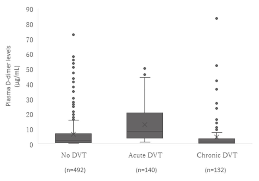
Figure 1 D-dimer levels in patients with and without DVT. The median D-dimer concentration (range) was 2.30 (0.2– 72.5) in the absence of DVT, 8.20( 1.1–50.0) in acute DVT, and 1.20( 0.3–83.4) in chronic DVT. Kruskal–Wallis rank sum test, p < 0.0001. DVT, Deep vein thrombosis.
IV. Discussion
The D-dimer test is useful because it can exclude DVT with high sensitivity 13). ELISA and latex agglutination methods are commercially available for measuring D-dimer and widely used in clinical laboratories, and the ELISA test is reported to be significantly more sensitive than latex agglutination in the DVT exclusion diagnosis 14). LG-DD is a reagent that measures D-dimer using the latex agglutination method, but the accuracy for low D-dimer concentrations is the same as that of ELISA 15). A comparison of the two methods using the transformation formula in the LG-DD package insert 9) showed a significant linear correlation.
In this study, 764 patients with venous thrombosis confirmed by US-echo were classified into three groups: no DVT, acute DVT, and chronic DVT. Then, LG-DD D-dimer values were compared among the patient groups. Patients with acute DVT showed significantly higher D-dimer levels compared with the no DVT and chronic DVT groups. This means that an increase in LG-DD D-dimer levels indicates new and active coagulation/fibrinolysis. Thus, the LG-DD D-dimer value distinguishes acute DVT from chronic and negative DVT without a need for US-echo.
Plasma levels of D-dimer in patients with malignant tumors increase because of ascites, pleural effusion retention, and increased coagulation/fibrinolysis 16)17). Post-operative D-dimer levels are increased by all three components of the Virchow triad, namely intravascular vessel wall damage, stasis of flow, and hypercoagulable status 18)19). Depending on these factors, the optimal cutoff of D-dimer in DVT exclusion diagnosis varies depending on the patient population.
Care must be taken in patients with underlying disorders that increase D-dimer levels. The specificity at a cutoff of 1.1 µg/mL, which is close to the normal range of D-dimer, was relatively low, reaching 15.4% in the malignant tumor patient group and 4.4% in the post-operative group. If the cutoff is near the normal range of D-dimer, the low specificity of exclusion diagnosis can lead to false positives. In contrast, the specificity of the optimal cutoff of 3.4 µg/mL for the malignant tumor patient group was improved to 55.8%, and the specificity at the optimal cutoff of 16.6 µg/mL for the post-operative group was significantly improved to 75.0%. However, the sensitivity of the post-operative group was significantly reduced to 58.6%. The cutoff for DVT exclusion diagnosis should result in high specificity while maintaining high NPV by employing a value that is appropriate for the given patient characteristics. It is not enough to just use the maximum Youden’s index of the ROC curve as a cutoff. Thus, we must collect data considering the timing of post-operative D-dimer measurements and select an effective way to determine an appropriate cutoff.
Aging is also one of the factors that increase D-dimer levels 7). In this study, an age-adjusted cutoff improved the specificity and NPV with high sensitivity compared with the cutoff of 1.1 µg/mL. The age-adjusted cutoff, calculated by multiplying age by 0.022, is simple and effective in eliminating the effects of age-related increases in D-dimer values.
Moreover, D-dimer values rapidly decrease with the administration of an anticoagulant. In our study, anticoagulants were administered to patients with acute DVT, and in most of these patients, D-dimer levels decreased rapidly and thrombus volume decreased or disappeared (data not shown). In addition, both acute DVT patients and chronic DVT patients had significantly lower D-dimer values than patients who did not use anticoagulants. The optimal cutoff value was slightly higher, and diagnostic accuracy was improved. Therefore, to diagnose DVT more accurately, it is necessary to evaluate D-dimer values before giving anticoagulants.
Table 1 Characteristics of 764 subjects who participated in this study. D-dimer levels. DVT, Deep vein thrombosis; Others, patients with various medical conditions that did not include malignant tumors, recovering from surgery, or pregnancy. *not applicable due to the small number of pregnant patients
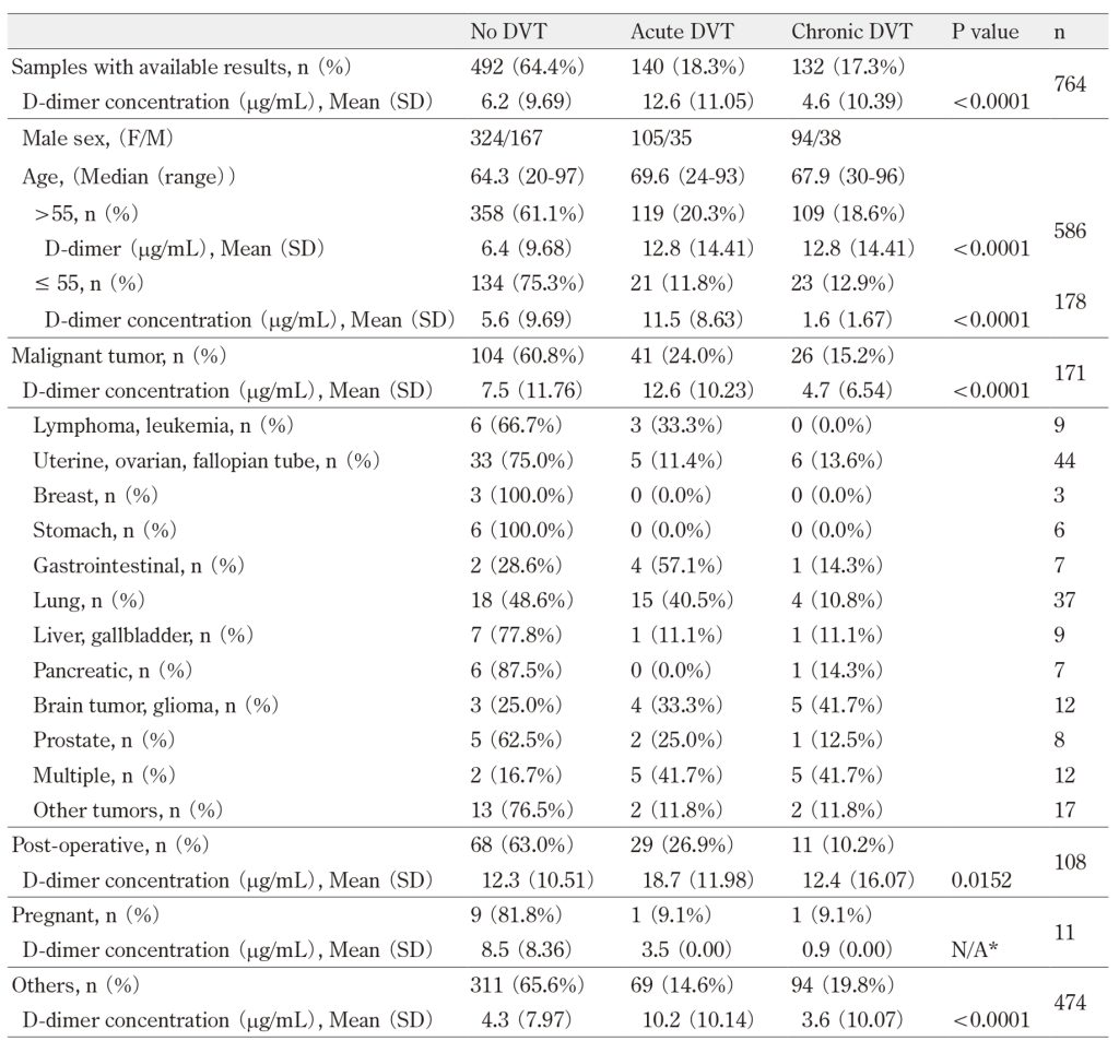
Table 2 Clinical performance of D-dimer assays by patient group. NPV, negative predictive value; PPV, positive
predictive value; LR-, negative likelihood ratio; LR+, positive likelihood ratio.
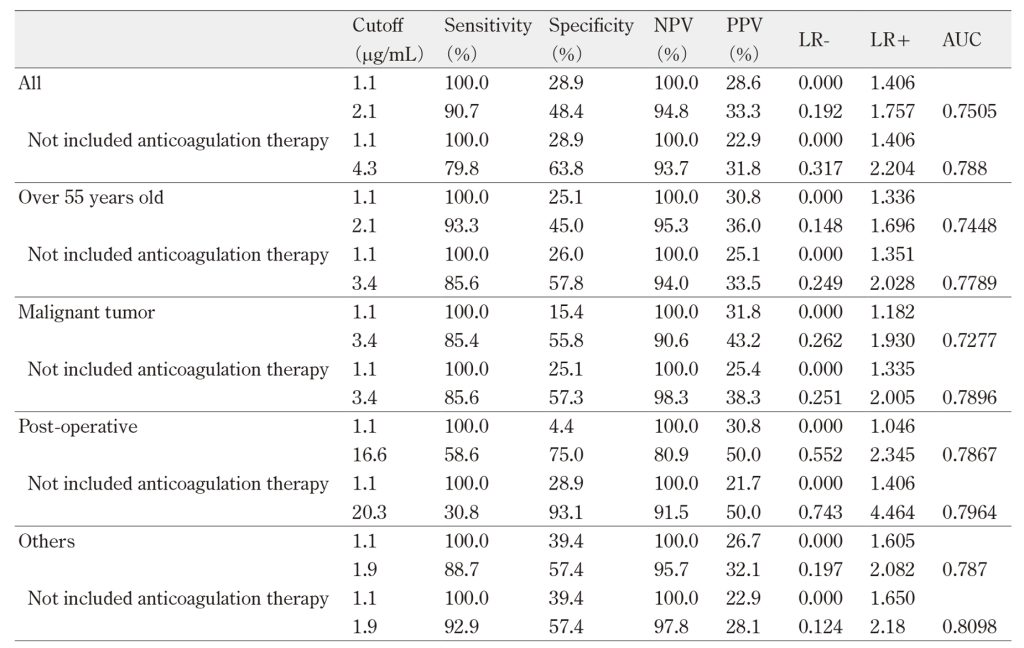
Table 3 Clinical performance of D-dimer assays in patients excluding those with malignant tumors, or who were
post-operative, or pregnant (n = 380). NPV, negative predictive value; PPV, positive predictive value;
LR-, negative likelihood ratio; LR+, positive likelihood ratio.

Table 4 D-dimer concentration with or without anticoagulants. DVT, deep vein thrombosis.

V. Conclusion
D-dimer levels are useful in DVT exclusion diagnosis, as measured by LG-DD. For more effective diagnoses of DVT exclusion, it is necessary to set an age-adjusted cutoff and higher cutoffs for post-operative patients and patients with malignant tumors.
Acknowledgment
This study was performed as a contract research program with LSI Medience Corporation, Tokyo, Japan.
Disclosure
The authors declare no conflicts of interest.
References
- Bates SM, Kearon C, Crowther, et al. A diagnostic strategy involving a quantitative latex D-dimer assay reliably excludes deep venous thrombosis. Ann Intern Med 2003; 138(10): 787-94.PubMed
- Nisio MD, Squizzato A, Rutjes AWS, et al. Diagnostic accuracy of D-dimer test for exclusion of venous thromboembolism: a systematic review. J Thromb Haemost 2007; 5(2): 296-304.PubMed
- Verma K, Legnani C, Palareti G. Cost-minimization analysis of venous thromboembolism diagnosis: Comparison of standalone imaging with a strategy incorporating D-dimer for exclusion of venous thromboembolism. Res Pract Thromb Haemost 2017; 1(1): 57-61.PubMed
- Kabrhel C, Courtney DM, Camargo CA, et al. Factors associated with positive D-dimer results in patients evaluated for pulmonary embolism. Acad Emerg Med 2010; 17(6): 589-97.PubMed
- JCS Joint Working Group. Guidelines for Diagnosis, Treatment and Prevention of Pulmonary Thromboembolism and Deep. Vein Thrombosis (JCS 2017). https://js-phlebology.jp/wp/wp-content/uploads/2019/03/JCS2017_ito_h.pdf
- Palareti G, Cosmi B, Legnani C, et al. D-dimer testing to determine the duration of anticoagulation therapy. N Engl J Med 2006; 355(17): 1780-9.PubMed
- Kearon C, Kahn SR. Long-term treatment of venous thromboembolism. Blood 2020; 135(5): 317-25.PubMed
- Farm M, Siddiqui AJ, Onelöv L, et al. Age-adjusted D-dimer cut-off leads to more efficient diagnosis of venous thromboembolism in the emergency department: a comparison of four assays. J Thromb Haemost 2018; 16(5): 866-75.PubMed
- Dempfle CE, Zips S, Ergül H, et al. The Fibrin Assay Comparison Trial (FACT): Evaluation of 23 Quantitative D-dimer Assays as Basis for the Development of D-dimer Calibrators. FACT study group. Thromb Haemost 2001; 85(4): 671-8.PubMed
- Sakata T. Standardization of markers related to thrombosis and haemostasis. Japanese Journal of Thrombosis and Hemostasis 2008; 19(4): 471-3.J-STAGE
- Johnson ED, Schell JC, Rodgers GM. The D-dimer assay. Am J Hematol 2019; 94(7): 833-9.PubMed
- Kanda Y. Investigation of the freely available easy-to-use software ‘EZR’ for medical statistics. Bone Marrow Transplant 2013; 48(3): 452-8.PubMed
- Wells PS, Anderson DR, Rodger M, et al. Evaluation of D-dimer in the diagnosis of suspected deep-vein thrombosis. N Engl J Med 2003; 349(13): 1227-35.PubMed
- Stein PD, Hull RD, Patel KC, et al. D-dimer for the exclusion of acute venous thrombosis and pulmonary embolism: a systematic review. Ann Intern Med 2004; 140(8): 589-602.PubMed
- Nagahama Y, Nozaki J, Sakurai G, et al. Clinical application of a new latex potometric immunoassay reagent, LPIA-GENESIS D-dimer, and its performance in patient-derived plasma samples. Int J Lab Hematol 2020; 42(3): 299-307.PubMed
- Falanga A, Marchetti M, Vignoli A. Coagulation and cancer: biological and clinical aspects. J Thromb Haemost 2013; 11(2): 223-33.PubMed
- Hisada Y, Mackman N. Cancer-associated pathways and biomarkers of venous thrombosis. Blood 2017; 130(13): 1499-506.PubMed
- Perka C. Preoperative versus postoperative initiation of thromboprophylaxis following major orthopedic surgery: safety and efficacy of postoperative administration supported by recent trials of new oral anticoagulants. Thromb J 2011; 9: 17.PubMed
- Joist JH. Hypercoagulability: introduction and perspective. Semin Thromb Hemost 1990; 16(2): 151-7.PubMed